. 2009 Sep 8;5(9):2486-2502.
doi: 10.1021/ct9001812.

Quantifying Correlations Between Allosteric Sites in Thermodynamic Ensembles

Affiliations

Quantifying Correlations Between Allosteric Sites in Thermodynamic Ensembles

Christopher L McClendon et al. J Chem Theory Comput. .

Abstract

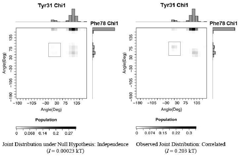
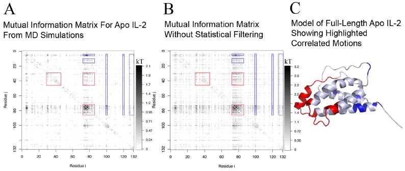
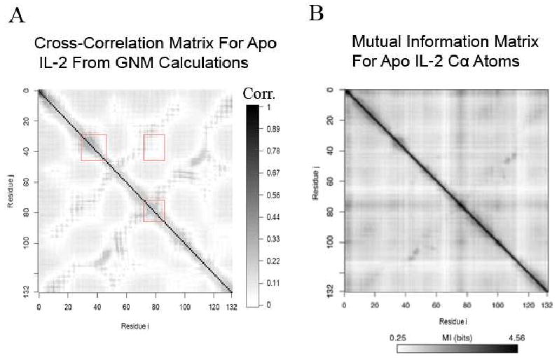
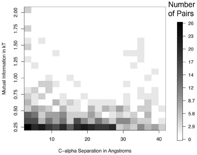
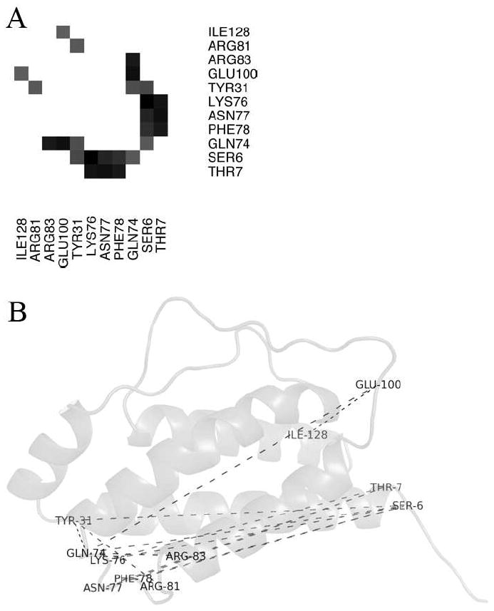
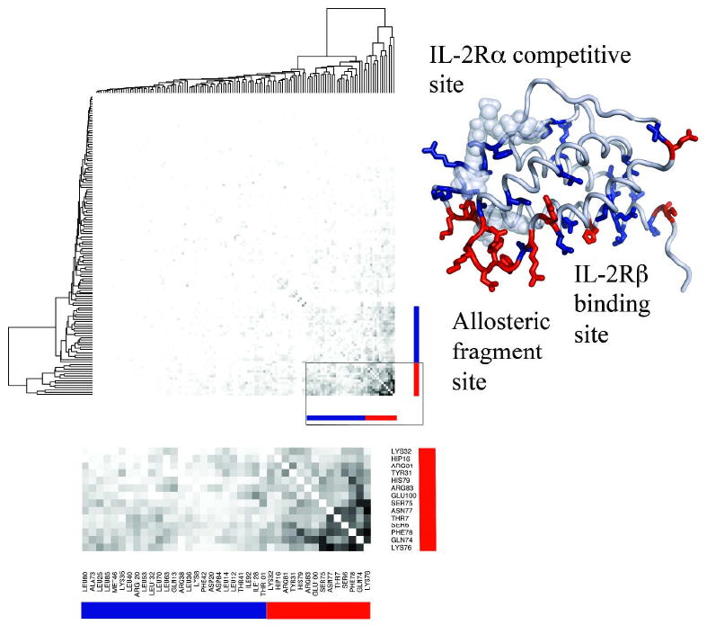
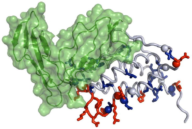
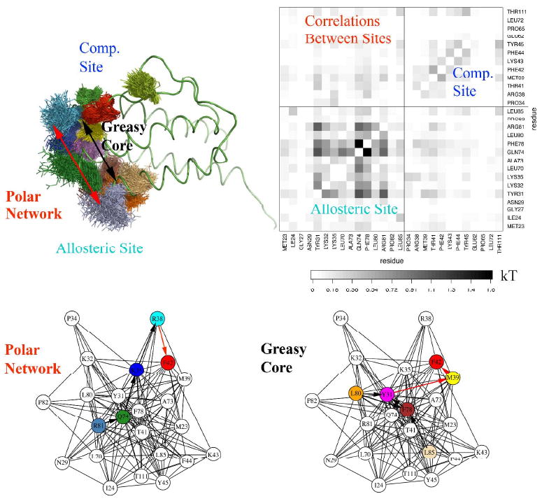
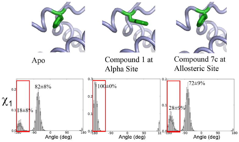
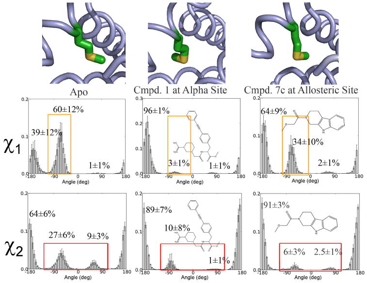
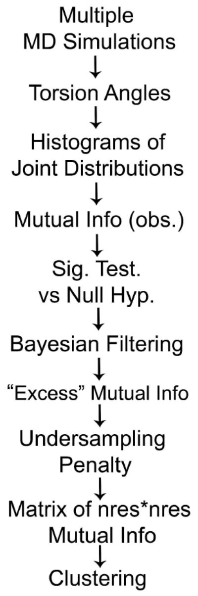
Allostery describes altered protein function at one site due to a perturbation at another site. One mechanism of allostery involves correlated motions, which can occur even in the absence of substantial conformational change. We present a novel method, "MutInf", to identify statistically significant correlated motions from equilibrium molecular dynamics simulations. Our approach analyzes both backbone and sidechain motions using internal coordinates to account for the gear-like twists that can take place even in the absence of the large conformational changes typical of traditional allosteric proteins. We quantify correlated motions using a mutual information metric, which we extend to incorporate data from multiple short simulations and to filter out correlations that are not statistically significant. Applying our approach to uncover mechanisms of cooperative small molecule binding in human interleukin-2, we identify clusters of correlated residues from 50 ns of molecular dynamics simulations. Interestingly, two of the clusters with the strongest correlations highlight known cooperative small-molecule binding sites and show substantial correlations between these sites. These cooperative binding sites on interleukin-2 are correlated not only through the hydrophobic core of the protein but also through a dynamic polar network of hydrogen bonding and electrostatic interactions. Since this approach identifies correlated conformations in an unbiased, statistically robust manner, it should be a useful tool for finding novel or "orphan" allosteric sites in proteins of biological and therapeutic importance.


Figures

Fig. 1
Fig. 1
Joint distributions of correlated torsions are different from what would be expected if they were independent. (A) Distributions of two χ1 torsion angles are shown along with the joint distribution expected if they were independent, i.e. the product of the marginal probabilities. (B) Distributions of the same two χ1 torsion angles are shown along with the observed joint distribution from molecular dynamics simulations. Grey boxes highlight a cross-peak with substantial height in the observed simulations (B) but with negligible height under the null hypothesis of independence (A).
Fig. 2
Fig. 2
Mutual Information captures significant correlations between residues in human interleukin-2. (A) Mutual information between residues' torsions computed using the present approach, with statistical filtering as detailed in Methods. (B) Same as in A but without any of the aforementioned statistical corrections. (C) The model of full-length human interleukin-2 used in the apo simulations, based on the crystal structure of apo IL-2 (PDB: 1M47). Residues surrounded by red boxes in A are colored red, while residues correlated to these that are surrounded by blue boxes in A are colored blue.
Fig. 3
Fig. 3
Comparison of pairwise, dynamical correlations between residues computed by alternative methods. (A) Absolute value of the cross-correlation matrix computed using the Gaussian Network Model. (B) Mutual Information between residues' Cα Cartesian coordinates using the approach of Lange and Grubmüller.
Fig. 4
Fig. 4
Most of the significant correlations are between distant residues. A 2-D histogram showing the number of pairs of correlated residues vs. Cα separation and mutual information value shows that the number and strength of correlated pairs decreases only modestly with distance. For clarity, only those pairs of correlations with a mutual information greater than 0.25 kT are shown.
Fig. 5
Fig. 5
Several distant residues are highly correlated. (A) Correlations greater than kT are shown only for IL-2 residues whose alpha carbons are separated by more than 5 Å. (B) Dashes connecting each pair of these correlated residues show long-rage correlations across the length of the helical bundle.
Fig. 6
Fig. 6
Hierarchical clustering of significant mutual information values identifies allosteric sites. A hierarchically-clustered heatmap shows clusters (top left) of residues with similar patterns of mutual information across IL-2 residues. A close-up view highlights numerous significant mutual information values between pairs of residues in two different clusters, red and blue. These red and blue clusters are highlighted in a model of IL-2's ternary complex (right). The strongest cluster (red sticks) chiefly involves a loop enclosing the allosteric fragment's binding site, and this cluster is correlated to a cluster (blue sticks) containing two protein binding sites, the IL-2Rα-receptor-binding/IL-2Rα-inhibitor-binding site and the IL-2Rβ-binding site. The two compound binding sites and the two protein-binding sites are directly correlated through the hydrophobic core (in the blue cluster), through a highly flexible loop (in the red cluster), and crosstalk between these elements, seen in a close-up view of the matrix (bottom).
Fig. 7
Fig. 7
Predicted couplings are consistent with regions perturbed upon IL-2Rα binding. Regions distant from the IL-2Rα receptor binding site that show substantial backbone chemical shift perturbations upon IL-2Rα binding roughly correspond to regions with residues whose conformations are correlated with residues in the IL-2Rα binding site (predominantly residues in the “blue” cluster in Fig. 6). Amides on IL-2 whose resonances shifted by more than three linewidths upon IL-2Rα binding or fell below 7% of the original intensity are shown as spheres. Residues from the “red and “blue” clusters shown in Fig. 6 are colored accordingly. IL-2 is as shown in cartoon and sticks as in Fig. 6, while IL-2Rα is shown in green.
Fig. 8
Fig. 8
Direct, pairwise correlations couple residues in the IL-2Rα-competitive site (at the IL-2:IL-2Rα interface) to residues in the allosteric fragment-binding site (near the IL-2:IL-2Rβ interface). (Top left) Apo IL-2 is shown in green ribbon while representative conformations of residues showing strong correlations within and between these sites are shown with lines. Overlap between clouds of residues' conformations suggest steric coupling, particularly in the greasy core, from Leu80 (orange) and Ler85 (tan) to Phe78 (brown), to Tyr31 (magenta), to Met39 (yellow), and to Phe42 (red). (Top right) A subset of the full matrix of pairwise correlations reveals direct correlations between residues in the two sites, with the labeled boxes showing correlations within the allosteric site, within the competitive site, and between these two sites. (Bottom) A force-directed network diagram for residues in these sites filtered for correlations of at least 0.05kT shows Phe78, Tyr31, Gln74, and Arg81 as central “hub” residues mediating correlations between the sites.
Fig. 9
Fig. 9
Compound binding to the allosteric site causes a population shift in the conformation of hot-spot residue Phe42 that favors binding compound at the IL-2Rα-competitive site. (Top) Conformations of Phe42 in apo and compound-bound crystal structures (PDB IDs 1M47, 1M48, and 1NBP, resp.). (Bottom) Histograms of Phe42's χ1 angle from MD simulations. Red boxes highlight the χ1 population selected by ligand binding at the competitive site.
Fig. 10
Fig. 10
Compound binding to the IL-2Rα site or to the allosteric site selects conformations of Met39 favorable for binding compound at the other site. (Top) Conformations of Met39 in apo and compound-bound crystal structures (PDB IDs 1M47, 1M48, and 1NBP, resp.). (Bottom) Histograms of Met39's χ1 and χ2 angles from MD simulations. Orange boxes in χ1 and red boxes in χ2 highlight populations suppressed in ligand-bound simulations.
Fig. 11
Fig. 11
Docking using Glide XP selects a holo-like conformation from an MD ensemble. (A, B) Plots of docking score vs. ligand RMSD to the forcefield-minimized co-crystal conformation show that the best-scoring docked poses from simulations with (B) but not without (A) allosteric compound bound had relatively low RMSD values. The best-scoring pose is circled. (C) Molecular dynamics snapshot from a simulation of IL-2 with bound allosteric fragment is shown with docked (yellow) vs. superimposed X-ray (magenta) conformations of a micromolar small molecule inhibitor of IL-2Rα binding. Though the absolute RMSD for this ligand is 2.9Å (1.6Å RMSD after fitting), it has a binding mode very similar to that of the crystal ligand.
Scheme 1
Scheme 1
A schematic of the MutInf approach for identifying correlated residue conformations shows how the observed mutual information is statistically filtered and corrected before being summed over residues pairs. The resulting matrix is then clustered as in a microarray experiment in order to identify groups of residues showing similar patterns of correlations.

Similar articles

Cited by

References

    1. Tsai CJ, del Sol A, Nussinov R. Allostery: Absence of a Change in Shape Does Not Imply that Allostery Is Not at Play. J Mol Biol. 2008;378(1):1–11. - PMC - PubMed
    1. Cui Q, Karplus M. Allostery and cooperativity revisited. Protein Sci. 2008;17(8):1295–1307. - PMC - PubMed
    1. Lindsley JE, Rutter J. Whence cometh the allosterome? Proc Natl Acad Sci U S A. 2006;103(28):10533–10535. - PMC - PubMed
    1. Gunasekaran K, Ma B, Nussinov R. Is allostery an intrinsic property of all dynamic proteins? Proteins: Struct, Funct, Bioinf. 2004;57(3):433–443. - PubMed
    1. Kuriyan J, Eisenberg D. The origin of protein interactions and allostery in colocalization. Nature. 2007;450(7172):983–990. - PubMed

LinkOut - more resources