. 2009 Oct;2(4):215-28, 228.e1-3.
doi: 10.1016/j.brs.2009.03.007.

Role of cortical cell type and morphology in subthreshold and suprathreshold uniform electric field stimulation in vitro

Affiliations

Role of cortical cell type and morphology in subthreshold and suprathreshold uniform electric field stimulation in vitro

Thomas Radman et al. Brain Stimul. 2009 Oct.

Abstract

Background: The neocortex is the most common target of subdural electrotherapy and noninvasive brain stimulation modalities, including transcranial magnetic stimulation (TMS) and transcranial current simulation (TCS). Specific neuronal elements targeted by cortical stimulation are considered to underlie therapeutic effects, but the exact cell type(s) affected by these methods remains poorly understood.

Objective: We determined whether neuronal morphology or cell type predicted responses to subthreshold and suprathreshold uniform electric fields.

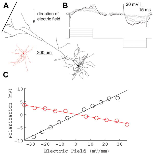
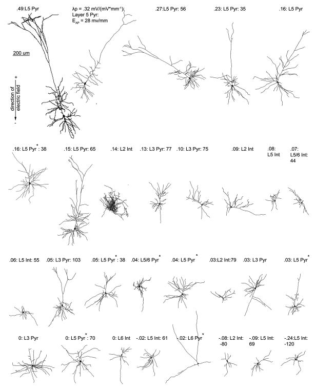
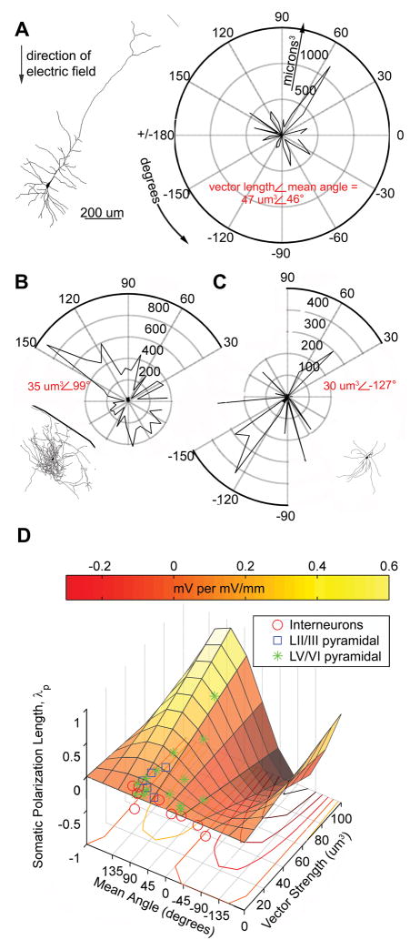
Methods: We characterized the effects of subthreshold and suprathreshold electrical stimulation on identified cortical neurons in vitro. Uniform electric fields were applied to rat motor cortex brain slices, while recording from interneurons and pyramidal cells across cortical layers, using a whole-cell patch clamp. Neuron morphology was reconstructed after intracellular dialysis of biocytin. Based solely on volume-weighted morphology, we developed a parsimonious model of neuronal soma polarization by subthreshold electric fields.

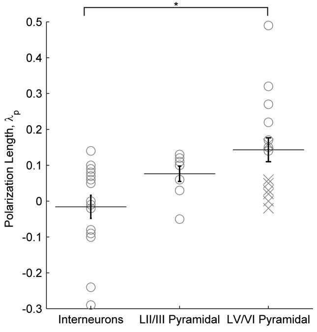
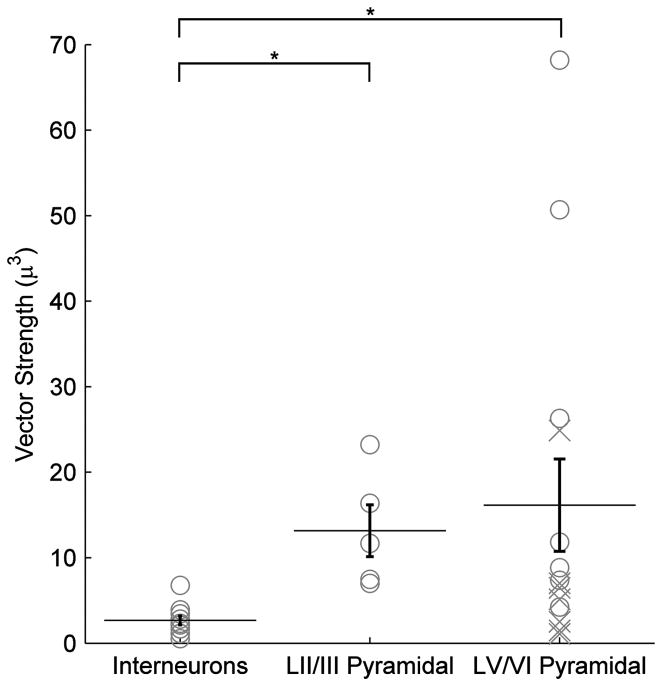
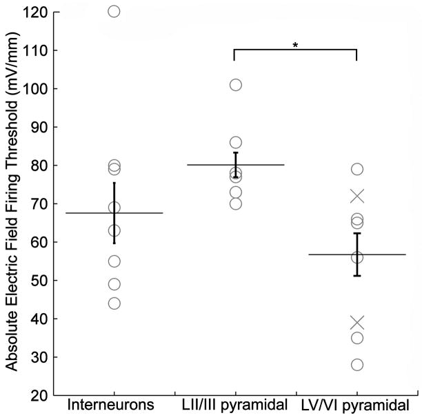
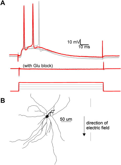
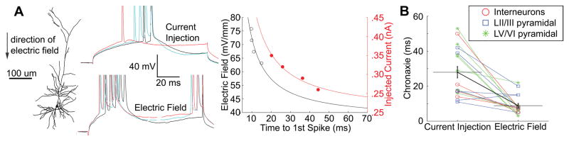
Results: We found that neuronal morphology correlated with somatic subthreshold polarization. Based on neuronal morphology, we predict layer V pyramidal neuronal soma to be individually the most sensitive to polarization by optimally oriented subthreshold fields. Suprathreshold electric field action potential threshold was shown to reflect both direct cell polarization and synaptic (network) activation. Layer V/VI neuron absolute electric field action potential thresholds were lower than layer II/III pyramidal neurons and interneurons. Compared with somatic current injection, electric fields promoted burst firing and modulated action potential firing times.

Conclusions: We present experimental data indicating that cortical neuron morphology relative to electric fields and cortical cell type are factors in determining sensitivity to sub- and supra-threshold brain stimulation.


Figures

Figure 1
Figure 1
Sub-threshold electric fields polarize cortical neuronal soma linearly. A, Example morphological reconstruction of a L5 pyramidal neuron (black), and L5 fast-spiking interneuron (red) B, Incrementing electric field steps of 5.8 mV/mm (bottom) linearly polarize cell soma (top). Reconstructions shown are from L5 regular spiking pyramidal neuron of A (top). C, Summary of the polarization per electric field for the neurons shown in A. The slope of the fitted line determines the sub-threshold field polarization sensitivity for each neuron. LV pyramidal neuron (black) = .27 mV*(mV/mm)−1, LV fast-spiking interneuron (red) = −.02 mV*(mV/mm)−1.
Figure 2
Figure 2
Cortical cell type polarization sensitivity. The polarization length, λp (mm), an indicator of mV or polarization per unit electric field applied (mV/mm), is shown according to cell type. Asterisk denotes significant difference (T-test) found between LV/VI pyramidal neurons and interneurons across layers. Points labeled as an “X” are neurons with cut dendritic trees that have still been included in all analyses.
Figure 3
Figure 3
Cortical neuron morphological reconstructions in order of electric field induced somatic polarization sensitivity. 3 items are listed for each cell, electric field induced somatic polarization length, λp (mm), an indicator of mV of polarization per unit electric field applied (mV/mm), layer, and cell type (pyramidal or interneuron); and if tested for that cell, electric field induced firing threshold. An asterisk next to the label for cell type denotes a neuron with a cut dendritic tree, that has still been included in all analyses.
Figure 4
Figure 4
Polar histogram coherence vector: Neuronal morphology predicts somatic polarization sensitivity to sub-threshold electric fields. A–C, Example tracings and corresponding volume-weighted polar histograms of (A) LV regular spiking pyramidal neuron with electric field The polarization length, λp (mm), an indicator of mV or polarization per unit electric field applied (mV/mm), = .32 mm, the polar histogram can be summarized by the variables: mean angle = 46° and vector length = 47 um3, representing the center of mass of the histogram; (B) LII fast spiking interneuron with polarization length = .14 mm, mean angle = 99° and vector length = 35 um3; and (C) LV fast spiking interneuron with polarization length = −.02 mm, mean angle = −127° and vector length = 30 um3. D, Summary plot of all neurons recorded and traced, with polar histogram coherence vectors as predictors of somatic polarization per electric field for each neuron. The colored plane is the statistically significant, best fit regression to the equation: polarization length = m*sine(mean angle)*vector length (p < .02, r2 = .41, n=30).
Figure 5
Figure 5
Cortical cell type vector lengths. The polar histogram (Fig. 4) summary variable vector length, is shown according to cell type. Asterisk denotes significant difference (T-test) found between as interneurons across layers and both LV/VI pyramidal neurons, and LII/III pyramidal neurons as well. Points labeled as an “X” are neurons with cut dendritic trees that have still been included in all analyses.
Figure 6
Figure 6
Cortical cell type electric field firing thresholds. The minimum absolute electric field firing threshold, in response to 100 ms incrementing electric field steps, is shown according to cell type. Asterisk denotes significant difference (T-test) found between LV/VI pyramidal neurons, and both LII/III pyramidal neurons as well as interneurons across layers.
Figure 7
Figure 7
Electric field induced EPSPs are reduced by bath application of CNQX and APV. A, Overlay of the response to electric field steps of increasing intensity. Top: Recorded intracellular voltage response to electric field steps of 51, 57, 63, and 70 (red trace) mV/mm. Note 63 and 70 mV/mm electric field steps induced action potentials. Middle: Voltage response to the same field intensities after 15 minute bath application of 20 μM CNQX and 50 μM APV. Bottom: Applied electric field waveforms. B, Tracing of L5 fast-spiking interneuron described in A.
Figure 8
Figure 8
Time to 1st spike-strength of stimulation chronaxie measurements are lower for electric field stimulation than somatic intracellular current injection. A, Example morphological reconstruction of a L5 intrinsic bursting pyramidal neuron with transmembrane polarization in response to successive steps of intracellular current injection (top), and electric field stimulation (bottom). Note modulation of firing pattern from regular spiking (top, current injection), to intrinsic bursting (bottom, field stimulation). Summary plot (right), of the time to first spike in response to electric field stimulation (left axis, black) and injected current (right axis, red). The solid lines are best fit curves to y=1/(time to first spike). B, Comparison of chronaxies, for current injection and electric field stimulation, for each recorded neuron. Statistically significant difference (p < .01) between stimulation methods for all cells.

Similar articles

Cited by

References

    1. Liebetanz D, et al. Anticonvulsant effects of transcranial direct-current stimulation (tDCS) in the rat cortical ramp model of focal epilepsy. Epilepsia. 2006;47(7):1216–24. - PubMed
    1. George MS, Lisanby SH, Sackeim HA. Transcranial magnetic stimulation: applications in neuropsychiatry. Arch Gen Psychiatry. 1999;56(4):300–11. - PubMed
    1. Avery DH, et al. A controlled study of repetitive transcranial magnetic stimulation in medication-resistant major depression. Biol Psychiatry. 2006;59(2):187–94. - PubMed
    1. Boggio PS, et al. Effects of transcranial direct current stimulation on working memory in patients with Parkinson’s disease. J Neurol Sci. 2006;249(1):31–8. - PubMed
    1. Webster BR, Celnik PA, Cohen LG. Noninvasive brain stimulation in stroke rehabilitation. NeuroRx. 2006;3(4):474–81. - PMC - PubMed

Publication types

MeSH terms