. 2009 Oct 21;1(3):3ra7.
doi: 10.1126/scitranslmed.3000139.

Radioprotection in normal tissue and delayed tumor growth by blockade of CD47 signaling

Affiliations

Radioprotection in normal tissue and delayed tumor growth by blockade of CD47 signaling

Justin B Maxhimer et al. Sci Transl Med. .

Abstract

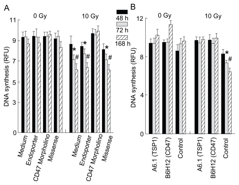
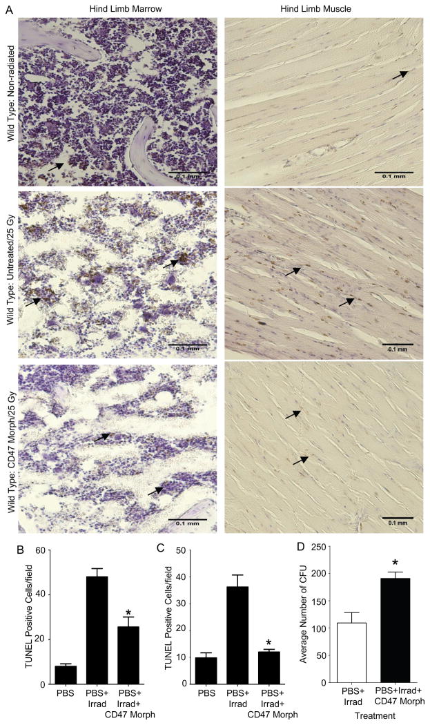
Radiation-induced damage of normal tissues restricts the therapeutic doses of ionizing radiation that can be delivered to tumors and thereby limits the effectiveness of radiotherapy. Thrombospondin-1 signaling through its cell surface receptor CD47 limits recovery from several types of stress, and mice lacking either gene are profoundly resistant to radiation injury. We describe strategies to protect normal tissues from radiation damage using CD47 or thrombospondin-1 antibodies, a CD47-binding peptide, or antisense suppression of CD47. A morpholino oligonucleotide targeting CD47 confers radioresistance to human endothelial cells in vitro and protects soft tissue, bone marrow, and tumor-associated leukocytes in irradiated mice. In contrast, CD47 suppression in mice bearing melanoma or squamous lung tumors prior to irradiation result in 89% and 71% smaller tumors, respectively. Thus, inhibiting CD47 signaling maintains the viability of normal tissues following irradiation while increasing the radiosensitivity of tumors.


Figures

Fig. 1
Fig. 1. Radioprotective activities of TSP1 and CD47 antibodies, a CD47 binding peptide from TSP1, and CD47 morpholino in human endothelial cells
HUVEC were treated with (A, E) TSP1 or (B, F) CD47 monoclonal antibodies (clone A6.1 or B6H12, respectively), (C, G) peptide 7N3, or (D) control peptide 604, and received the indicated doses of radiation 24 h after addition of the antibodies. Cell (mitochondrial) viability was measured 48 h after irradiation by MTS reduction (A–D), and cell proliferation was examined by BrdU incorporation into newly synthesized DNA (E–G). (H) Knockdown of CD47 by pretreatment of HUVEC for 48 h with a specific morpholino complexed with Endoporter vehicle protected against cell death as assessed by MTS reduction after a radiation dose of 40 Gy when compared to control groups (non-treated, mismatched morpholino complexed with Endoporter, and Endoporter vehicle control) (*P <0.05, #P < 0.01). (I) Cells treated with a morpholino to suppress the TSP1 receptor CD36 under the same conditions demonstrated no radioprotection. Experiments were repeated 3 times, and results are presented as the mean optical density ± SD.
Fig. 2
Fig. 2. Antibodies to TSP1 and CD47 and antisense suppression of CD47 maintain proliferation of irradiated human endothelial cells
HUVEC were treated with (A) CD47 morpholino or (B) TSP1 or CD47 antibodies and exposed to 10 Gy irradiation 48 hours later. DNA synthesis was assessed at the indicated times by BrdU incorporation, and relative fluorescence (RFU) is presented as the mean ± SD (*P < 0.05, #p <0.01). Experiments were repeated 3 times.
Fig. 3
Fig. 3. The radioprotective effect of CD47 suppression is independent of NO/cGMP signaling
HUVEC were treated with the indicated doses of the slow releasing NO donor DETA/NO (A) or membrane permeable cGMP analog, 8-Bromo cGMP (B). Cell viability was measured by MTS reduction 48 hours after 40 Gy irradiation. (C) HUVEC or HUVEC treated with CD47 morpholino were incubated with the indicated concentrations of nitric oxide synthase inhibitor L-NAME prior to irradiation, and cell viability was measured by MTS reduction.
Fig. 4
Fig. 4. CD47 suppression with an antisense morpholino minimizes tissue and vascular damage from radiation injury
Age and sex matched C57BL/6 wild type mice underwent a one-time treatment of the right hindlimb with either a CD47 morpholino oligonucleotide (10 μM in 750 μl PBS) or 750 μl of PBS only injected into the muscle and soft tissues and received 25 Gy irradiation to the treated hindlimb 48 hours later. (A) After 8 weeks, a representative image shows signs of fibrotic contractures only in the untreated irradiated hind limb. (B) Tissue necrosis grading scores for 8 animals were calculated weekly by assessing alopecia, ulceration, and desquamation and are presented as mean ± SD. (C) Fibrosis was assessed at 8 weeks by measuring hindlimb extension and is presented as mean ± SD (*P < 0.05). (D) C57BL/6 wild type mice were treated by direct intra-muscular injection of the right hindlimb with the CD47 morpholino (10 μM in 750 μl PBS, upper panels) or sterile PBS alone (lower panels) 48 hours prior to 25 Gy irradiation of both hindlimbs in each mouse. Eight weeks after radiation, right hindlimb blood flow responses to DEA/NO challenge (100 nmol/gram body weight via rectal instillation) were assessed via Laser Doppler imaging under 1.5% isoflurane inhalation anesthesia at a core temperature of 36.5° C. Representative images show a greater increase in blood flow after 25 min. in the right hindlimb of a morpholino treated mouse (arrows in upper panels) compared to the corresponding limb of a vehicle treated mouse (lower panels). (E) Blood flow velocity measurements (Flux) at the indicated time points were integrated over the area of the treated hindlimbs of 6 mice in each group and normalized as a percent of the integrated baseline flux in the same limb of each mouse. Results are presented as the mean ± SD.
Fig. 5
Fig. 5. Suppression of CD47 prevents radiation-induced apoptosis in muscle and bone marrow
(A) Un-irradiated control hindlimbs (upper panels) and hindlimbs irradiated at a dose of 25 Gy (middle and bottom panels) from age and sex matched C57BL/6 wild type mice were prepared for paraffin-embedded tissue sections 24 h post-radiation and stained for apoptotic nuclei by the TUNEL method (brown nuclear staining). Mouse hindlimbs injected with the CD47 morpholino (bottom panels) 48 h prior to radiation were also prepared in the same manner 24 h post-radiation. Apoptosis is inferred by intranuclear staining of muscle and bone marrow cells. Images were acquired using a 20x objective (B) Bone marrow apoptosis was quantified for 5 mice in each group and is presented as the average number of TUNEL positive cells in 3 tissue sections per animal (*P<0.005). (C) Apoptosis in muscle was quantified for 5 mice in each group and is presented as the average number of TUNEL positive cells in 3 tissue sections per animal (*P<0.001). (D) Assessment of colony forming units of haematopoietic progenitor cell derived from bone marrow cells of mice treated in the same manner as explained above. Data represent mean ± SEM of 6 mice per treatment group (*P<0.01).
Fig. 6
Fig. 6. CD47 suppression enhances re-growth delay in irradiated tumors but increases radioresistance of melanoma cells in vitro.
(A) C57BL/6 mice were injected with 1×10⁶ B16 mouse melanoma cells into their right hindlimbs. Five days later, the mice were randomized into 4 groups (12 mice in each group) that received injections of CD47 morpholino (10 μM) in sterile PBS, PBS, or no injection. Two groups receiving injections were then subjected to radiation (10 Gy) to the affected hindlimb 48 h later. The other two groups served as controls. Tumor volume was measured every 6 days for 30 days. Results are presented as the mean ± SD of 12 mice in each treatment group and are representative of 3 independent studies. (B) The same experimental design was used to assess radiation delay for SCC VII squamous cell lung carcinoma tumors (SCC, 1.5×10⁵ cells). Results are presented as the mean ± SD of 10 mice (C3H) in each treatment group and are representative of 3 independent studies. (C) Mice bearing SCC-VII tumors as in (B) were treated by local intramuscular and subcutaneous (SC) injections of PBS or CD47 morpholino or by intraperitoneal (IP) injection of CD47 morpholino, irradiated at 10 Gy, and tumor growth was assessed at the indicated times. (D) B16 melanoma cells plated on 96-well plates in standard growth medium were treated with peptide 7N3 or CD47 morpholino and received a radiation dose of 10 Gy. Cell (mitochondrial) viability was measured 48 h after radiation using the MTS assay (*P <0.05, #P < 0.01).
Fig. 7
Fig. 7. Increased fibrotic response, viable leukocyte infiltrates, and macrophage infiltration in tumors with suppressed CD47 expression
C57BL/6 mice were injected with 1×10⁶ B16 mouse melanoma cells into their hindlimbs. Treatment consisted of two groups in which the hindlimbs were treated with either 150 μl of PBS or 150 μl of CD47 morpholino (10 μM) 48 hours prior to radiation (10 Gy). The control group consisted of tumor without radiation. 24 h post-radiation the mice were sacrificed, and paraffin embedded tissue sections of the hindlimbs were prepared for analysis. (A) H&E stained sections of hindlimbs and tumors. (B) Detection of cells undergoing apoptosis by the TUNEL method. Apoptotic cells are indicated by brown nuclear staining (arrows). (C) Immunohistochemical staining for murine macrophage surface marker CD68. Dark brown staining within the tumor indicates macrophage infiltration. Images were acquired using either a 4x or 10x objective. (D) Quantitative analysis of apoptotic tumor-associated cells by TUNEL staining, n= 5 in each group, *P < 0.0001) (E) Quantitative analysis of tumor-associated macrophages detected by CD68-immunostaining. (63)

Similar articles

Cited by

References

    1. Nair CK, Parida DK, Nomura T. Radioprotectors in radiotherapy. J Radiat Res (Tokyo) 2001;42:21–37. - PubMed
    1. Emami B, et al. Tolerance of normal tissue to therapeutic irradiation. Int J Radiat Oncol Biol Phys. 1991;21:109–122. - PubMed
    1. Stone HB, Coleman CN, Anscher MS, McBride WH. Effects of radiation on normal tissue: consequences and mechanisms. Lancet Oncol. 2003;4:529–536. - PubMed
    1. Duchstein S, Gademann G, Peters B. Early and late effects of local high dose radiotherapy of the brain on memory and attention. Strahlenther Onkol. 2003;179:441–451. - PubMed
    1. Coleman CN. Radiation oncology--linking technology and biology in the treatment of cancer. Acta Oncol. 2002;41:6–13. - PubMed