. 2009 Dec 23;1(12):12ra23.
doi: 10.1126/scitranslmed.3000540.

Measurement and clinical monitoring of human lymphocyte clonality by massively parallel VDJ pyrosequencing

Affiliations

Measurement and clinical monitoring of human lymphocyte clonality by massively parallel VDJ pyrosequencing

Scott D Boyd et al. Sci Transl Med. .

Abstract

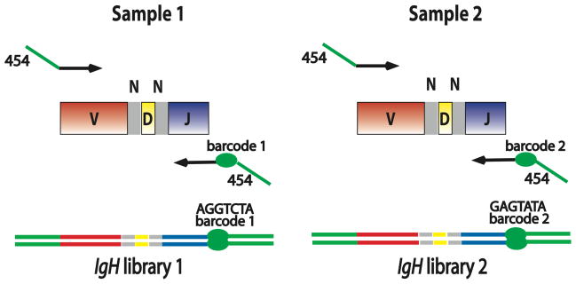
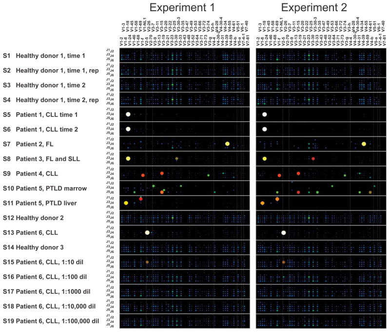
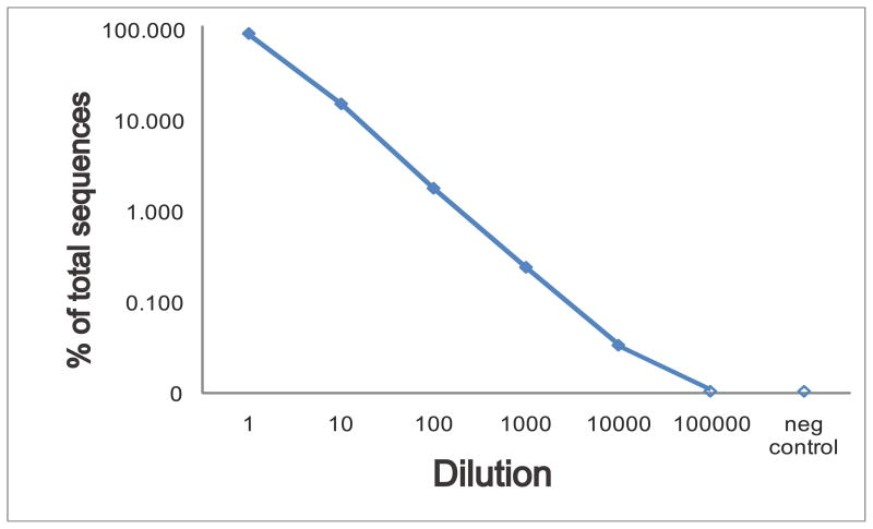
The complex repertoire of immune receptors generated by B and T cells enables recognition of diverse threats to the host organism. In this work, we show that massively parallel DNA sequencing of rearranged immune receptor loci can provide direct detection and tracking of immune diversity and expanded clonal lymphocyte populations in physiological and pathological contexts. DNA was isolated from blood and tissue samples, a series of redundant primers was used to amplify diverse DNA rearrangements, and the resulting mixtures of barcoded amplicons were sequenced using long-read ultra deep sequencing. Individual DNA molecules were then characterized on the basis of DNA segments that had been joined to make a functional (or nonfunctional) immune effector. Current experimental designs can accommodate up to 150 samples in a single sequence run, with the depth of sequencing sufficient to identify stable and dynamic aspects of the immune repertoire in both normal and diseased circumstances. These data provide a high-resolution picture of immune spectra in normal individuals and in patients with hematological malignancies, illuminating, in the latter case, both the initial behavior of clonal tumor populations and the later suppression or re-emergence of such populations after treatment.


Conflict of interest statement

Conflicts of Interest

Boyd, Marshall, Merker, Maniar, Zhang, Sahaf, Jones, Nadeau, Nguyen, Miklos, Zehnder, Fire: None Simen, Hanczaruk, and Egholm are employees of 454 Life Sciences, A Roche Company

Figures

Fig. 1
Fig. 1. Barcoded PCR amplicons for multiplexed IgH sequencing
PCR primers used for preparing barcoded amplicons for high-throughput sequencing were designed using the FR2 IgH V gene segment family primers and the common IgH J segment primer from the BIOMED-2 consortium (19). Additional sequences required for emulsion PCR and pyrosequencing were added (indicated in green) at the 5′ end of the IgH-specific primers. In addition, a 6-, 7-, or 10-nucleotide sequence barcode was designed into the modified IgH J primer to identify the sample from which the PCR amplicons were derived. In the specimens analyzed using the 454 Titanium sequencer, an additional 10-nucleotide sample barcode was incorporated into the multiplexed IgH V gene segment primers used for amplification (Supplementary Table 1). Lines with arrowheads indicate PCR primers. Green segments: primer sequences needed for 454 sequencing protocol; red segments: V gene segment sequence; grey segments: non-templated N base sequences; yellow segments: D gene segment sequence; blue segments: J gene segment sequence; green ellipse: sample-specific barcode enabling pooling of IgH libraries for multiplexed sequencing. Sample 1 and Sample 2 could represent DNA template from any two clinical specimens, or independent DNA template aliquots from the same specimen.
Fig. 2
Fig. 2. Immunoglobulin heavy chain V and J gene segment usage in healthy peripheral blood, oligoclonal or indeterminate specimens, and lymphoid malignancy specimens
Barcoded IgH rearrangement libraries were PCR-amplified from genomic DNA of human specimens, pooled, and characterized by high-throughput pyrosequencing. Experiments 1 and 2 were independent experimental replicates beginning with different aliquots of the template DNA from each specimen. Each wide row represents the IgH sequences identified in a single sample. Samples (S1–S19) are labeled in the far-left column in the figure. The x-axis (across the top of the panels) indicates the V gene segment used in the receptor, and the y-axis (the column at the left of the panels) within each wide row represents the J gene segments used. The size and color of the circle at a given point indicates what proportion of all sequences in the sample used that particular combination of V and J gene segments. Sequences in which V, D or J segments or junctions could not be unambiguously assigned were filtered prior to generation of these plots. Rep: replicate sequence pool PCR amplified from an independent aliquot of template DNA; CLL: chronic lymphocytic leukemia; FL: follicular lymphoma; SLL: small lymphocytic lymphoma; PTLD: post-transplant lymphoproliferative disorder.
Fig. 3
Fig. 3. Titration of a chronic lymphocytic leukemia clonal sample into healthy peripheral blood
Pooled barcoded IgH library sequencing was carried out on a series of 10-fold dilutions of a chronic lymphocytic leukemia blood sample (sample 13) into a healthy control blood sample (sample 14), to evaluate the sensitivity and linearity of high-throughput sequencing for detection of a known clonal sequence. The percentage of sequences matching the chronic lymphocytic leukemia clone in each diluted specimen is plotted on a log scale, with zero indicating that no sequences were detected. The counts of clonal sequences in each sample were as follows: CLL sample, 7805 clonal of 8612 total; healthy blood control, 0 clonal of 7518 total; 1:10 dilution, 2095 clonal of 13,717 total; 1:100 dilution, 156 clonal of 8674 total; 1:1000 dilution, 23 clonal of 9471 total; 1:10,000 dilution, 3 clonal of 8895 total; 1:100,000 dilution, 0 clonal of 6940 total. The negative control is the healthy donor blood sample used for diluting the clonal CLL sample. A second experiment measuring fewer sequences from independent PCR amplifications from the same samples detected the following number of clonal sequences in each sample: CLL sample, 422 clonal of 566 total; healthy blood control, 0 clonal of 270 total; 1:10 dilution, 189 clonal of 665 total; 1:100 dilution, 11 clonal of 230 total; 1:1000 dilution, 0 clonal of 344 total; 1:10,000 dilution, 0 clonal of 329 total; 1:100,000 dilution, 0 clonal of 208 total.

Similar articles

Cited by

References

    1. Schatz DG. Antigen receptor genes and the evolution of a recombinase. Semin Immunol. 2004;16:245–256. - PubMed
    1. Davis MM, Bjorkman PJ. T-cell antigen receptor genes and T-cell recognition. Nature. 1988;334:395–402. - PubMed
    1. Arstila TP, Casrouge A, Baron V, Even J, Kanellopoulos J, Kourilsky P. A direct estimate of the human alphabeta T cell receptor diversity. Science. 1999;286:958–961. - PubMed
    1. Brezinschek HP, Brezinschek RI, Lipsky PE. Analysis of the heavy chain repertoire of human peripheral B cells using single-cell polymerase chain reaction. J Immunol. 1995;155:190–202. - PubMed
    1. Lim A, Luderschmidt S, Weidinger A, Schnopp C, Ring J, Hein R, Ollert M, Mempel M. The IgE repertoire in PBMCs of atopic patients is characterized by individual rearrangements without variable region of the heavy immunoglobulin chain bias. J Allergy Clin Immunol. 2007;120:696–706. - PubMed

Publication types

MeSH terms

LinkOut - more resources