. 2010 Feb 9;4(2):e598.
doi: 10.1371/journal.pntd.0000598.

Temporal expression of chemokines dictates the hepatic inflammatory infiltrate in a murine model of schistosomiasis

Affiliations

Temporal expression of chemokines dictates the hepatic inflammatory infiltrate in a murine model of schistosomiasis

Melissa L Burke et al. PLoS Negl Trop Dis. .

Abstract

Schistosomiasis continues to be an important cause of parasitic morbidity and mortality world-wide. Determining the molecular mechanisms regulating the development of granulomas and fibrosis will be essential for understanding how schistosome antigens interact with the host environment. We report here the first whole genome microarray analysis of the murine liver during the progression of Schistosoma japonicum egg-induced granuloma formation and hepatic fibrosis. Our results reveal a distinct temporal relationship between the expression of chemokine subsets and the recruitment of cells to the infected liver. Genes up-regulated earlier in the response included T- and B-cell chemoattractants, reflecting the early recruitment of these cells illustrated by flow cytometry. The later phases of the response corresponded with peak recruitment of eosinophils, neutrophils, macrophages and myofibroblasts/hepatic stellate cells (HSCs) and the expression of chemokines with activity for these cells including CCL11 (eotaxin 1), members of the Monocyte-chemoattractant protein family (CCL7, CCL8, CCL12) and the Hepatic Stellate Cell/Fibrocyte chemoattractant CXCL1. Peak expression of macrophage chemoattractants (CCL6, CXCL14) and markers of alternatively activated macrophages (e.g. Retnla) during this later phase provides further evidence of a role for these cells in schistosome-induced pathology. Additionally, we demonstrate that CCL7 immunolocalises to the fibrotic zone of granulomas. Furthermore, striking up-regulation of neutrophil markers and the localisation of neutrophils and the neutrophil chemokine S100A8 to fibrotic areas suggest the involvement of neutrophils in S. japonicum-induced hepatic fibrosis. These results further our understanding of the immunopathogenic and, especially, chemokine signalling pathways that regulate the development of S. japonicum-induced granulomas and fibrosis and may provide correlative insight into the pathogenesis of other chronic inflammatory diseases of the liver where fibrosis is a common feature.


Conflict of interest statement

The authors have declared that no competing interests exist.

Figures

Figure 1
Figure 1. Parasite burden and granulofibrotic pathology.
Infected mice harboured a mean of 5 worm pairs (A). Schistosome eggs were first observed in the liver at 4 weeks p.i and hepatic egg burden increased significantly thereafter (1-Way ANOVA, p≤0.05) (B). Granuloma volume (C) and collagen staining for hepatic fibrosis (D) increased significantly from 4 weeks p.i, reaching 51% and 28% total liver volume at 7 weeks p.i, respectively (1-Way ANOVA, p≤0.01). Values represent mean values from 4 mice pooled for microarray analysis ±1SD.* p≤0.05, ** p≤0.01, ns = not significant compared with uninfected liver unless otherwise indicated.
Figure 2
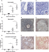
Figure 2. Changes in the distribution of eosinophils, neutrophils and myofibroblasts/HSCs in the murine liver during S. japonicum infection.
The number of eosinophils in the liver increased significantly from 4 weeks p.i (A). Eosinophils (arrows) were first observed within small inflammatory infiltrates adjacent to blood vessels (V) (B: Giemsa ×400) and, later, within granulomas associated with schistosome eggs (C: Giemsa ×400). Neutrophils were first observed in the liver at 6 weeks p.i in small inflammatory foci and their numbers increased significantly thereafter (D). At 7 weeks p.i. neutrophils occurred in the centre of established granulomas adjacent to schistosome eggs (E: Leder stain ×200) and at the periphery of more fibrotic granulomas (F: Leder stain ×100). α-SMA staining for myofibroblasts/HSCs increased significantly in infected compared with uninfected liver at 7 weeks p.i (G) and was localised to the fibrotic zone of granulomas (H: α-SMA staining, ×100; I: Picosirius red staining of the same granuloma, ×100). Scale bar equals 100 µm. Values represent means of 4 samples pooled for microarray analysis ±1SD. *p≤0.05, **p≤0.01, ***p≤0.001 compared with uninfected liver unless otherwise indicated.
Figure 3
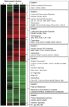
Figure 3. S. japonicum infection is associated with temporal expression of genes with distinct biological functions.
Hierarchical clustering identified seven distinct clusters representing genes that were consistently up-regulated during infection (Cluster 3); up-regulated to a greater extent later in infection (Cluster 2 and Cluster 4); up-regulated to a greater extent earlier in infection (Cluster 5 and Cluster 7); and down-regulated during infection (Cluster 1 and Cluster 6). Prominent biological functions, signalling pathways or chemokines associated with each of these clusters are listed in the boxed text. Gene expression is represented as a heat map with relatively unchanged genes coloured black, down regulated genes coloured green and up-regulated genes coloured red.
Figure 4
Figure 4. Temporal changes in the cellular composition of the S. japonicum-infected murine liver revealed by flow cytometry.
Flow cytometry showed an increase in the number of CD4+ T-cells (CD4+CD3+) (A), CD8+ T-cells (CD8+CD3+) (B), and B-cells (CD19+) (C) in the liver at 4 weeks p.i, but there was a decline in the numbers of these cells thereafter. The number of macrophages in the liver (D) was significantly increased at 4 weeks p.i. compared with tissue from uninfected mice and the number of these cells continued to rise over time peaking at 6-7 weeks p.i. Values represent means ± 1SD. One mouse from the 4 and 6 weeks groups harboured no adult worms (i.e. non-infected) and was excluded from these analyses (n = 4). n = 5 all other groups. * p≤0.05, ** p≤0.01, ***p≤0.001 compared with uninfected liver unless otherwise indicated.
Figure 5
Figure 5. S100A8 and CCL7 localise to areas of known neutrophil accumulation and the fibrotic zone of S. japonicum induced granulomas, respectively.
S100A8 positive (pink) cells occurred sporadically in the livers of uninfected mice (A; arrows; ×100), in the central region of established granulomas at 6 weeks p.i. (B; ×100) and at the periphery of granulomas (C; ×100) adjacent to fibrotic areas identified by Pico Sirius Red staining for collagen (pink) (D; ×100) at 7 weeks p.i. Whereas, uninfected liver was negative, CCL7 (Red-brown/arrows) was significantly increased at 7 weeks p.i. and localised to areas of fibrosis (E; ×100, & F; ×200), as identified by Pico Sirius Red staining for collagen (G; ×200), resembling the distribution of α-SMA staining (H; ×200). Scale bar equals 100 µm.
Figure 6
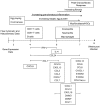
Figure 6. Summary of chemokines with distinct functions that exhibited peak expression during different phases of the S. japonicum-induced granulofibrotic response.
Chemokines up-regulated earlier were predominantly T- and B-cell chemoattractants. Chemokines up-regulated in the mid-late stages of infection are associated with HSC recruitment and activation. Chemokines with peak expression late in infection were predominantly macrophage and neutrophil chemoattractants. The activity of these chemokines was reflected in the temporal recruitment of T-cells, B-cells, eosinophils, neutrophils, macrophages and myofibroblasts/HSCs to the granulomatous liver.

Similar articles

Cited by

References

    1. King CH. Schistosomiasis japonica: The DALYs recaptured. PLoS Negl Trop Dis. 2008;2:e203. - PMC - PubMed
    1. Burke ML, Jones MK, Gobert GN, Li YS, Ellis MK, et al. Immunopathogenesis of human schistosomiasis. Parasite Immunol. 2009;31:163–176. - PubMed
    1. Cheever AW. Comparison of pathologic changes in mammalian hosts infected with Schistosoma mansoni, S. japonicum and S. haematobium. Mem Inst Oswaldo Cruz. 1987;82(Suppl 4):39–45. - PubMed
    1. Hsu SY, Hsu HF, Davis JR, Lust GL. Comparative studies on the lesions caused by eggs of Schistosoma japonicum and Schistosoma mansoni in livers of albino mice and rhesus monkeys. Ann Trop Med Parasitol. 1972;66:89–97. - PubMed
    1. Warren KS, Grove DI, Pelley RP. The Schistosoma japonicum egg granuloma. II. Cellular composition, granuloma size, and immunologic concomitants. Am J Trop Med Hyg. 1978;27:271–275. - PubMed

Publication types