

Telomere shortening sensitizes cancer cells to selected cytotoxic agents: in vitro and in vivo studies and putative mechanisms
- PMID: 20161752
- PMCID: PMC2817744
- DOI: 10.1371/journal.pone.0009132
Telomere shortening sensitizes cancer cells to selected cytotoxic agents: in vitro and in vivo studies and putative mechanisms
Abstract
Background: Telomere/telomerase system has been recently recognized as an attractive target for anticancer therapy. Telomerase inhibition results in tumor regression and increased sensitivity to various cytotoxic drugs. However, it has not been fully established yet whether the mediator of these effects is telomerase inhibition per se or telomere shortening resulting from inhibition of telomerase activity. In addition, the characteristics and mechanisms of sensitization to cytotoxic drugs caused by telomerase inhibition has not been elucidated in a systematic manner.
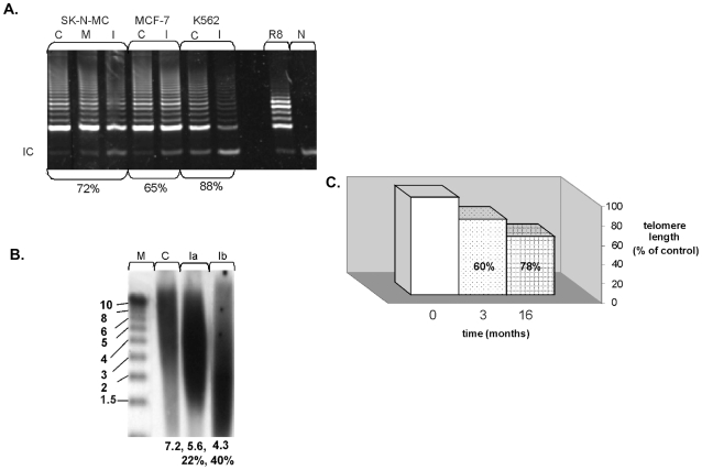
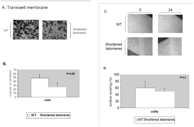
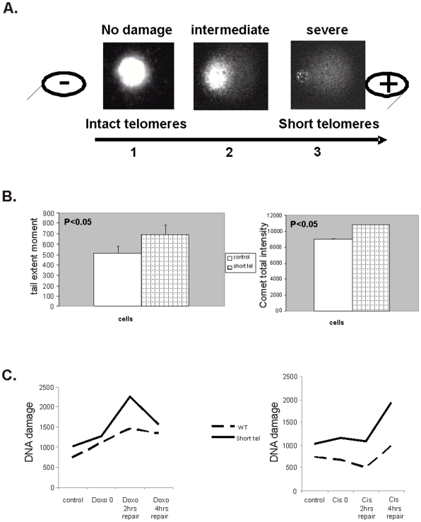
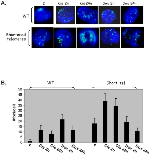
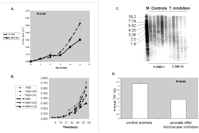
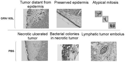
Methodology/principal findings: In this study we characterized the relative importance of telomerase inhibition versus telomere shortening in cancer cells. Sensitization of cancer cells to cytotoxic drugs was achieved by telomere shortening in a length dependent manner and not by telomerase inhibition per se. In our system this sensitization was related to the mechanism of action of the cytotoxic drug. In addition, telomere shortening affected also other cancer cell functions such as migration. Telomere shortening induced DNA damage whose repair was impaired after administration of cisplatinum while doxorubicin or vincristine did not affect the DNA repair. These findings were verified also in in vivo mouse model. The putative explanation underlying the phenotype induced by telomere shortening may be related to changes in expression of various microRNAs triggered by telomere shortening.
Conclusions/significance: To our best knowledge this is the first study characterizing the relative impact of telomerase inhibition and telomere shortening on several aspects of cancer cell phenotype, especially related to sensitivity to cytotoxic drugs and its putative mechanisms. The microRNA changes in cancer cells upon telomere shortening are novel information. These findings may facilitate the development of telomere based approaches in treatment of cancer.
Conflict of interest statement
Figures

Similar articles
-
Telomerase antagonists GRN163 and GRN163L inhibit tumor growth and increase chemosensitivity of human hepatoma.Hepatology. 2005 Nov;42(5):1127-36. doi: 10.1002/hep.20822. Hepatology. 2005. PMID: 16114043
-
Telomerase inhibitor imetelstat has preclinical activity across the spectrum of non-small cell lung cancer oncogenotypes in a telomere length dependent manner.Oncotarget. 2016 May 31;7(22):31639-51. doi: 10.18632/oncotarget.9335. Oncotarget. 2016. PMID: 27192120 Free PMC article.
-
Thymoquinone induces telomere shortening, DNA damage and apoptosis in human glioblastoma cells.PLoS One. 2010 Aug 12;5(8):e12124. doi: 10.1371/journal.pone.0012124. PLoS One. 2010. PMID: 20711342 Free PMC article.
-
How to inhibit telomerase activity for cancer therapy.Curr Med Chem Anticancer Agents. 2002 Sep;2(5):613-26. doi: 10.2174/1568011023353796. Curr Med Chem Anticancer Agents. 2002. PMID: 12678728 Review.
-
Targeting human telomerase in cancer therapy.Curr Med Chem Anticancer Agents. 2002 Sep;2(5):577-87. doi: 10.2174/1568011023353822. Curr Med Chem Anticancer Agents. 2002. PMID: 12678725 Review.
Cited by
-
The combined use of known antiviral reverse transcriptase inhibitors AZT and DDI induce anticancer effects at low concentrations.Neoplasia. 2012 Jan;14(1):44-53. doi: 10.1593/neo.11426. Neoplasia. 2012. PMID: 22355273 Free PMC article.
-
Keeping those telomeres short! an innovative intratumoral long-term drug delivery system.J Cancer Res Clin Oncol. 2015 Jan;141(1):23-34. doi: 10.1007/s00432-014-1747-7. Epub 2014 Jul 30. J Cancer Res Clin Oncol. 2015. PMID: 25073436 Free PMC article.
-
Effect of targeted silencing of hTERT mRNA by lentivirus-mediated siRNA on A549 lung cancer cells in vitro.Mol Biol Rep. 2013 Jan;40(1):605-16. doi: 10.1007/s11033-012-2099-5. Epub 2012 Oct 9. Mol Biol Rep. 2013. PMID: 23054018
-
Oligonucleotides Targeting Telomeres and Telomerase in Cancer.Molecules. 2018 Sep 5;23(9):2267. doi: 10.3390/molecules23092267. Molecules. 2018. PMID: 30189661 Free PMC article. Review.
-
Exosomal telomerase transcripts reprogram the microRNA transcriptome profile of fibroblasts and partially contribute to CAF formation.Sci Rep. 2022 Sep 30;12(1):16415. doi: 10.1038/s41598-022-20186-8. Sci Rep. 2022. PMID: 36180493 Free PMC article.
References
-
- Palm W, de Lange T. How Shelterin Protects Mammalian Telomeres. Annu Rev Genet. 2008;42:301–34. - PubMed
-
- Shay JW, Wright WE. Telomeres and telomerase: implications for cancer and aging. Radiat Res. 2001;155:188–93. - PubMed
-
- Collins K, Mitchell JR. Telomerase in the human organism. Oncogene. 2002;21:564–79. - PubMed
-
- Lavelle F, Riou JF, Laoui A, Mailliet P. Telomerase: a therapeutic target for the third millennium? Crit Rev Oncol Hematol. 2000;34:111–26. - PubMed
-
- Cong Y, Shay JW. Actions of human telomerase beyond telomeres. Cell Res. 2008;18:725–32. - PubMed
Publication types
MeSH terms
Substances
LinkOut - more resources
Full Text Sources
Other Literature Sources
