

Comparison of breast cancer to healthy control tissue discovers novel markers with potential for prognosis and early detection
- PMID: 20161755
- PMCID: PMC2817747
- DOI: 10.1371/journal.pone.0009122
Comparison of breast cancer to healthy control tissue discovers novel markers with potential for prognosis and early detection
Erratum in
- PLoS One. 2010;5(4). doi: 10.1371/annotation/632c5ae8-271b-4d19-8509-dc3b2eefe6a4 doi: 10.1371/annotation/632c5ae8-271b-4d19-8509-dc3b2eefe6a4
Abstract
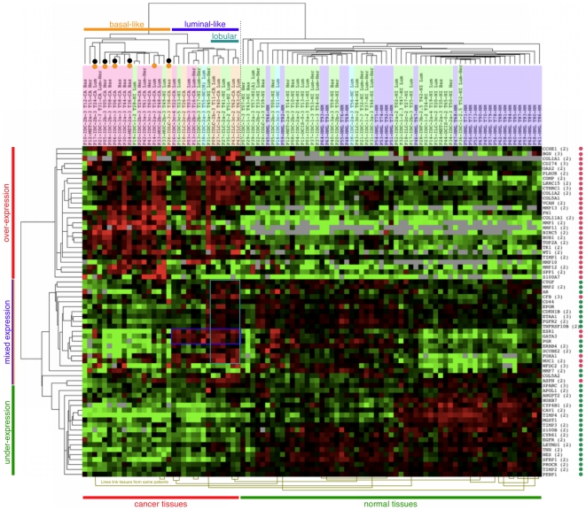
This study was initiated to identify biomarkers with potential value for the early detection of poor-outcome breast cancer. Two sets of well-characterized tissues were utilized: one from breast cancer patients with favorable vs. poor outcome and the other from healthy women undergoing reduction mammaplasty. Over 46 differentially expressed genes were identified from a large list of potential targets by a) mining publicly available expression data (identifying 134 genes for quantitative PCR) and b) utilizing a commercial PCR array. Three genes show elevated expression in cancers with poor outcome and low expression in all other tissues, warranting further investigation as potential blood markers for early detection of cancers with poor outcome. Twelve genes showed lower expression in cancers with poor outcome than in cancers with favorable outcome but no differential expression between aggressive cancers and most healthy controls. These genes are more likely to be useful as prognostic tissue markers than as serum markers for early detection of aggressive disease. As a secondary finding was that, when histologically normal breast tissue was removed from a distant site in a breast with cancer, 7 of 38 specimens displayed a cancer-like expression profile, while the remaining 31 were genetically similar to the reduction mammaplasty control group. This finding suggests that some regions of ipsilateral histologically 'normal' breast tissue are predisposed to becoming malignant and that normal-appearing tissue with malignant signature might warrant treatment to prevent new primary tumors.
Conflict of interest statement
Figures
References
-
- Schummer M, Ng W, Bumgarner R, Nelson P, Schummer B, et al. Comparative hybridization of an array of 21,500 ovarian cDNAs for the discovery of genes overexpressed in ovarian carcinomas. Gene. 1999;238:375–385. - PubMed
-
- Pepe MS, Etzioni R, Feng Z, Potter JD, Thompson ML, et al. Phases of biomarker development for early detection of cancer. J Natl Cancer Inst. 2001;93:1054–1061. - PubMed
-
- Hellström I, Raycraft J, Hayden-Ledbetter M, Ledbetter J, Schummer M, et al. The HE4 (WFDC2) protein is a biomarker for ovarian carcinoma. Cancer Res. 2003;63:3695–3700. - PubMed
-
- Duffy MJ. Serum tumor markers in breast cancer: are they of clinical value? Clin Chem. 2006;52:345–351. - PubMed
Publication types
MeSH terms
Substances
Grants and funding
LinkOut - more resources
Full Text Sources
Medical
Research Materials
