. 2010 Feb 9;5(2):e9135.
doi: 10.1371/journal.pone.0009135.

Phenylbutyric acid rescues endoplasmic reticulum stress-induced suppression of APP proteolysis and prevents apoptosis in neuronal cells

Affiliations

Phenylbutyric acid rescues endoplasmic reticulum stress-induced suppression of APP proteolysis and prevents apoptosis in neuronal cells

Jesse C Wiley et al. PLoS One. .

Abstract

Background: The familial and sporadic forms of Alzheimer's disease (AD) have an identical pathology with a severe disparity in the time of onset [1]. The pathological similarity suggests that epigenetic processes may phenocopy the Familial Alzheimer's disease (FAD) mutations within sporadic AD. Numerous groups have demonstrated that FAD mutations in presenilin result in 'loss of function' of gamma-secretase mediated APP cleavage [2], [3], [4], [5]. Accordingly, ER stress is prominent within the pathologically impacted brain regions in AD patients [6] and is reported to inhibit APP trafficking through the secretory pathway [7], [8]. As the maturation of APP and the cleaving secretases requires trafficking through the secretory pathway [9], [10], [11], we hypothesized that ER stress may block trafficking requisite for normal levels of APP cleavage and that the small molecular chaperone 4-phenylbutyrate (PBA) may rescue the proteolytic deficit.

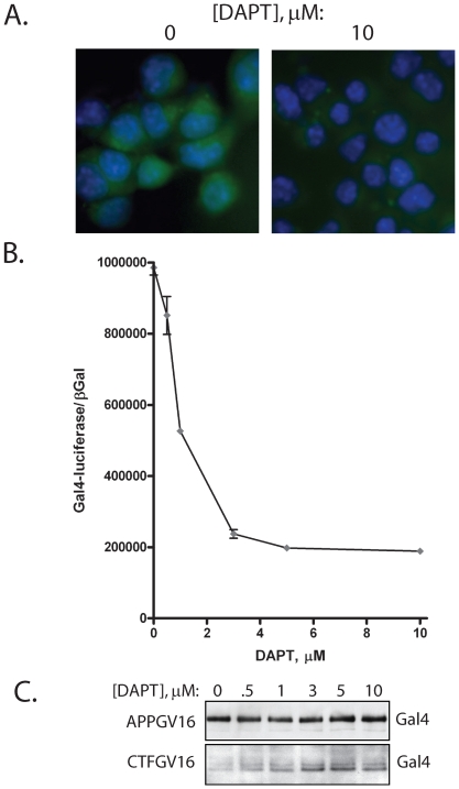
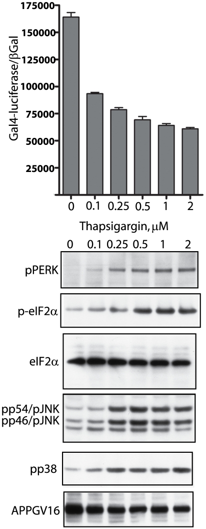
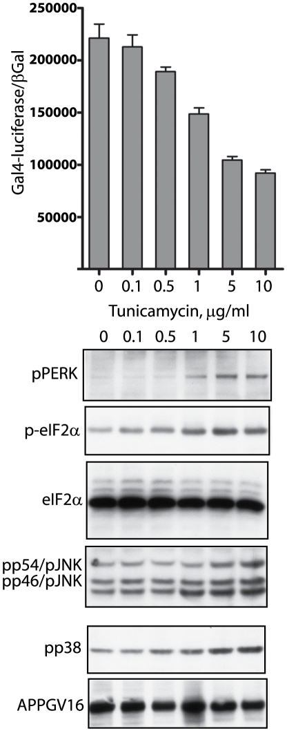
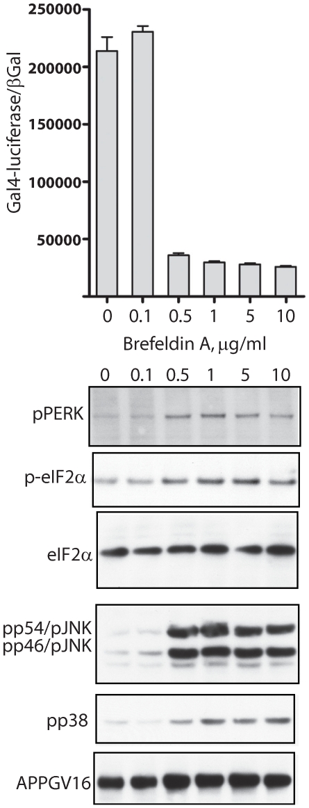
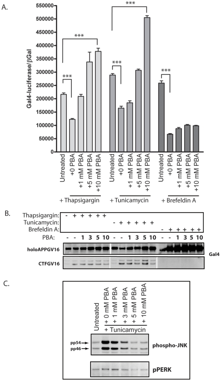
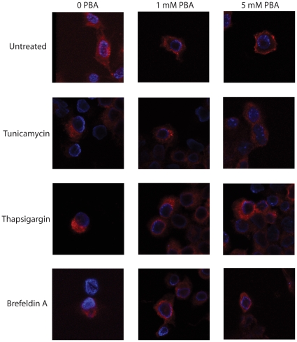
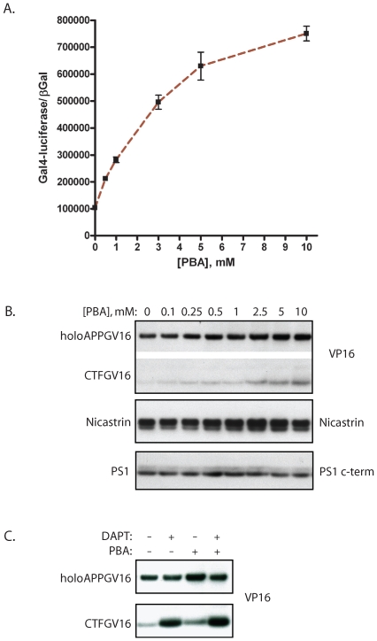
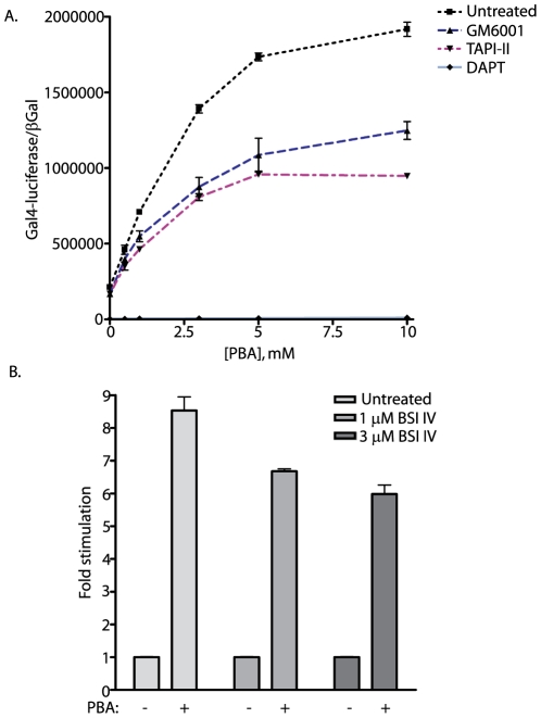
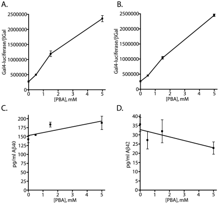
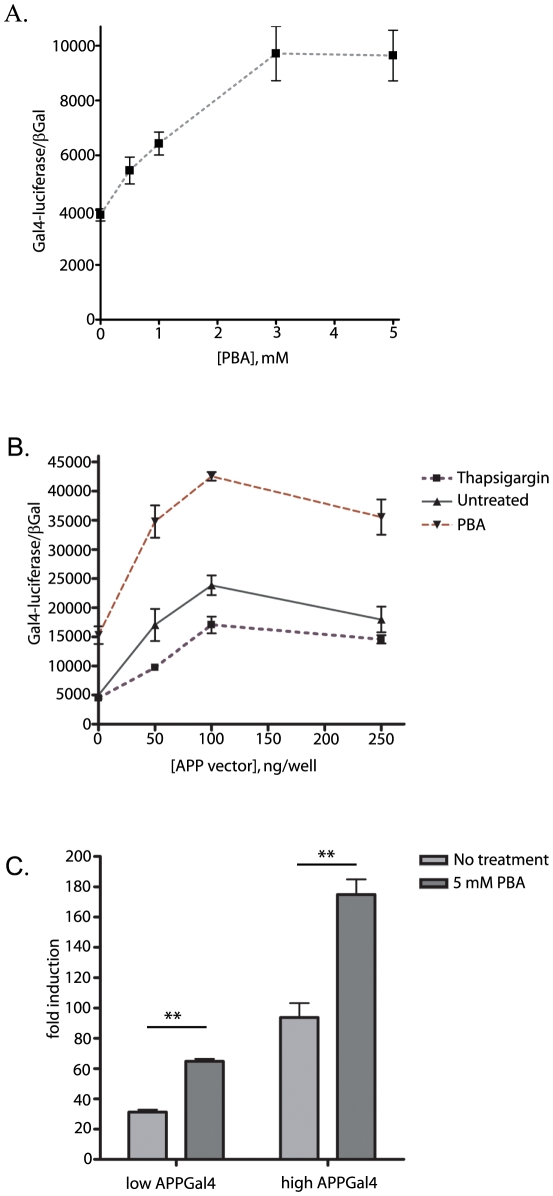
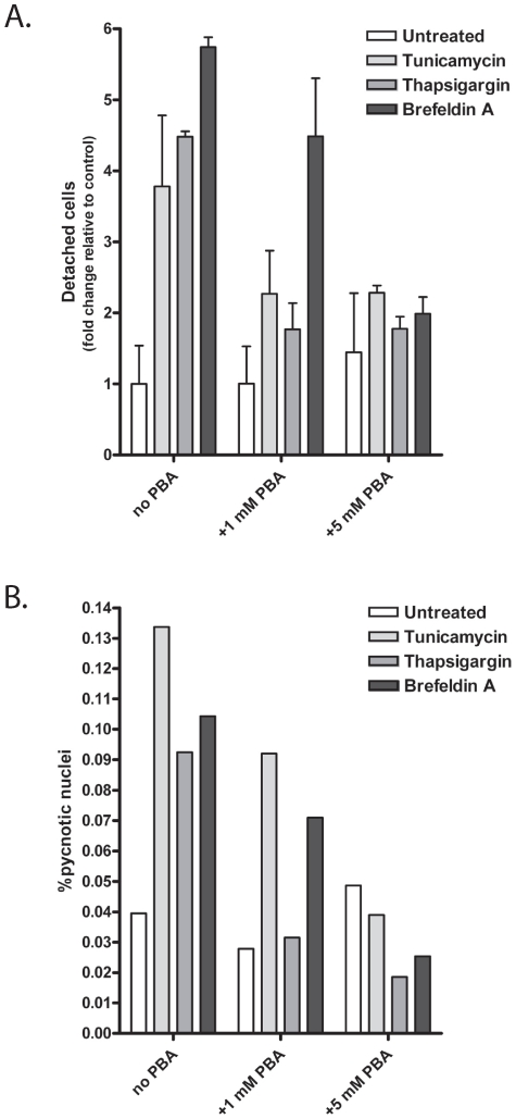
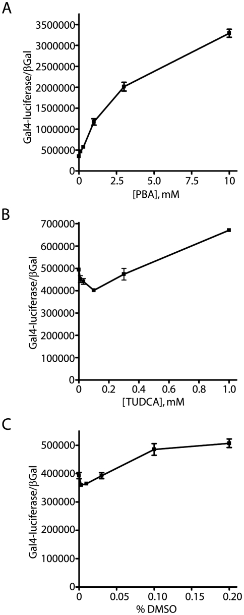
Methodology/principal findings: The APP-Gal4VP16/Gal4-reporter screen was stably incorporated into neuroblastoma cells in order to assay gamma-secretase mediated APP proteolysis under normal and pharmacologically induced ER stress conditions. Three unrelated pharmacological agents (tunicamycin, thapsigargin and brefeldin A) all repressed APP proteolysis in parallel with activation of unfolded protein response (UPR) signaling-a biochemical marker of ER stress. Co-treatment of the gamma-secretase reporter cells with PBA blocked the repressive effects of tunicamycin and thapsigargin upon APP proteolysis, UPR activation, and apoptosis. In unstressed cells, PBA stimulated gamma-secretase mediated cleavage of APP by 8-10 fold, in the absence of any significant effects upon amyloid production, by promoting APP trafficking through the secretory pathway and the stimulation of the non-pathogenic alpha/gamma-cleavage.

Conclusions/significance: ER stress represses gamma-secretase mediated APP proteolysis, which replicates some of the proteolytic deficits associated with the FAD mutations. The small molecular chaperone PBA can reverse ER stress induced effects upon APP proteolysis, trafficking and cellular viability. Pharmaceutical agents, such as PBA, that stimulate alpha/gamma-cleavage of APP by modifying intracellular trafficking should be explored as AD therapeutics.


Conflict of interest statement

Competing Interests: The authors have declared that no competing interests exist.

Figures

Figure 1
Figure 1. γ-secretase dependence of the APPGV16/Gal4-luciferaseEGFP/EF1- NLS-βGal reporter cells.
The N2a EF1-APPGV16/Gal4-luciferaseEGFP/EF1-NLS-βGal stable cells (NAG cells) have γ-secretase dependent EGFP expression, as DAPT eliminates the majority of EGFP fluorescence (A). The Gal4-luciferase moiety of the reporter is also γ-secretase dependent as titrated DAPT diminishes luciferase activity (B). The decrease in luciferase activity correlates with elevated CTFGV16 protein levels across the DAPT titration (C).
Figure 2
Figure 2. Thapsigargin inhibits APP proteolysis and induces ER stress UPR signaling.
Increasing doses of thapsigargin inhibits γ-secretase cleavage of APPGV16 (top graph). Titrated doses of thapsigargin induced graded increases in UPR stress signaling through phospho-PERK (pPERK blot) and phospho-eIF2α (p-eIF2 α), beginning at 0.1 µM and reaching a plateau at 0.5 µM thapsigargin. The increases in UPR signaling correlate with the levels of phospho-p38 (pp38 blot) and phospho-JNK (pp54/JNK, pp46JNK blot), as maximal stimulation is observed in the same concentration range. Total protein expression levels remain unchanged, as eIF2α and APPGV16 protein levels were consistent across treatment conditions.
Figure 3
Figure 3. Tunicamycin inhibits APP proteolysis and induces ER stress UPR signaling.
The NAG cells were treated with titrated doses of the N-glycosylation inhibitor tunicamycin, which inhibits γ-secretase cleavage of APPGV16 (top graph). Increasing doses of tunicamycin induced elevated levels UPR activation, demonstrated by elevated levels of phospho-PERK (pPERK blot) and phospho-eIF2α (p-eIF2 α) levels. Tunicamycin elevated phospho-p38 (pp38 blot) and phospho-JNK (pp54/JNK, pp46JNK blot) levels in parallel with increased UPR signaling and decreased APPGV16 proteolysis.
Figure 4
Figure 4. Brefeldin A inhibits APP proteolysis and induces ER stress UPR signaling.
Brefeldin A inhibited γ-secretase cleavage of APPGV16 (top graph). Western blots show that 0.5 µg/ml Brefeldin A induces activation of PERK (pPERK blot) and eIF2α (p-eIF2 α). Increases in phospho-p38 (pp38 blot) and phospho-JNK (pp54/JNK, pp46JNK blot) levels are also observed at the same concentration of BFA which fosters increased UPR signaling and diminished γ-secretase dependent APPGV16 cleavage.
Figure 5
Figure 5. Small molecular chaperone 4-phenylbutyric acid (PBA) rescues APP proteolysis from thapsigargin and tunicamycin treatment.
NAG cells either remained untreated, or were treated with thapsigargin (0.25 µg/ml), tunicamycin (5 µg/ml), or brefeldin A (5 µg/ml) alone, or in conjunction with titrated values of PBA, for 24 hours. Gal4-luciferase measures were normalized to constitutive NLS-βGal expression. Treatment with each stress inducing compound resulted in significant decreases in APPGV16 proteolysis (p<0.0002, unpaired t-test) (A). Co-treatment with PBA rescued the decrease in APPGV16 proteolysis, promoting cleavage beyond that observed in untreated cells in the presence of thapsigargin or tunicamycin (p<0.0001, t-test). PBA had no significant effect upon APPGV16 proteolysis in the NAG cells treated with brefeldin A. The effects of the stress inducing compounds and PBA upon APPGV16 and CTFGV16 protein levels were examined in parallel experiments (B). Thapsigargin and tunicamycin treatment resulted in elevated CTFGV16 levels. Co-treatment with PBA resulted in minor decreases in CTFGV16 levels by the titration endpoint. No CTFGV16 proteolytic fragments were observed in the brefeldin A treated NAG cells. PBA inhibited UPR signaling (phospho-JNK and phospho-PERK) induced by 5 µg/ml tunicamycin (C).
Figure 6
Figure 6. PBA stimulates trafficking out of the intracellular organelles.
NAG cells were untreated, or treated with thapsigargin (0.25 µM), tunicamycin (5 µg/ml), or brefeldin A (5 µg/ml) in the presence or absence of PBA for 24 hours. The cells were stained with the VP16 antibody (red) and counterstained with Hoescht (blue) to localize the nuclei. Confocal imaging was performed to examine protein localization. In untreated cells, APPGV16 localized throughout the cytosol with slight peri-nuclear aggregations observed, consistent with the ER localization of de novo membrane proteins. PBA treatment promoted migration of APPGV16 toward the plasma membrane (top row). In tunicamycin, thapsigargin and brefeldin A treated cells, APPGV16 localized to the ER-like perinuclear region (left column, lower three images). Upon the addition of 1 mM or 5 mM PBA, APPGV16 localization shifted away from the nucleus in tunicamycin and thapsigargin treated NAG cells. In contrast, PBA had no effect upon the localization of APPGV16 in the brefeldin A treated cells.
Figure 7
Figure 7. PBA stimulates secretase-mediated APP cleavage.
The NAG cells were treated with titrated concentrations of PBA for 24 hours. PBA elicited elevated levels of Gal4-reporter reporter transactivation in a concentration dependent fashion (p<0.0001; student t-test 0 versus 10 mM PBA) (A). PBA effects upon APPGV16, PS1 and Nicastrin expression levels were examined in the NAG cells 24 hours post-treatment. APP-Gal4VP16 levels increased slightly across the PBA titration. The CTFGV16 proteolytic fragment, whose molecular weight corresponds to the α-cleavage product of APPGV16, increased substantially across the PBA titration (B, top blot). In contrast, no increases in Nicastrin or PS1 levels were observed (B, middle and bottom blots). CTFGV16 increased in NAG cells treated with PBA and DAPT. Co-treatment with DAPT and PBA increased CTFGV16 protein levels in an additive manner, consistent with PBA stimulating CTF-GV16 production (C).
Figure 8
Figure 8. PBA stimulation of APPGV16 requires secretase processing.
In order to determine whether PBA stimulation of APPGV16 is dependent upon proteolytic processing, NAG cells were treated with titrated levels of PBA in the presence or absence of specific secretase inhibitor:. 20 µM GM6001 (broad-spectrum metalloprotease inhibitor), 50 µM TAPI-II (α-secretase inhibitor), or 20 µM DAPT (γ-secretase inhibitor). PBA stimulated a statistically significant 9-fold increase in APP proteolysis (p<0.0001; student t-test). However, co-treatment with GM6001 or TAPI-II decreased the fold-stimulation by approximately half (p<0.0001; two-way ANOVA). DAPT significantly reduced the levels of activity and eliminated the stimulatory effect of PBA (p<0.0001; two-way ANOVA). The β-secretase contribution to PBA enhanced APP proteolysis was examined using two different concentrations of beta-secretase inhibitor (BSI) IV (B). PBA stimulated an 8–9 fold increase in normalized Gal4-luciferase activity. BSI IV decreased Gal4-luciferase activity by less than 25% (p<0.01; student t-test) (B). These data suggest that α-secretase plays a more significant role than β-secretase in PBA enhanced APP proteolysis.
Figure 9
Figure 9. Amyloid biogenesis (Aβ40 and Aβ42) is unaffected by the PBA mediated stimulation in AICD production.
The NAG cells were treated with titrated values of PBA (0, 0.5, 1.5, and 5 mM). A two-part assay measured γ-secretase dependent AICD production (A and B) and secreted amyloid biogenesis (C and D) from the same samples. The Gal4-reporter assays were performed with the cell lysate, while the media was assayed for species specific amyloid concentrations. PBA stimulated γ-secretase mediated proteolysis approximately ten-fold (A and B) in parallel assays to the ELISA measures for Aβ40 (C) and Aβ42 (D). Each concentration step in the PBA titration induced a statistically significant increase in Gal4-reporter activity ([PBA] shift: 0 to 0.5 mM, p<0.0003; 0.5 mM to 1.5 mM, p<0.0002; 1.5 mM to 5 mM, p<0.0001; analysis performed with the values in (A)). In contrast, there was no statistically significant change in either Aβ40 or Aβ42 levels. Aβ40 levels increased slightly from 142 picograms/ml to 188 picograms/ml, with a p-value of 0.09. In contrast, Aβ42 levels decreased from 35.6 picograms/ml to 22.7 picograms/ml, with a p-value of 0.06. In total, Aβ40 levels increased by 32.3 percent and Aβ42 levels decreased 36.05 percent—neither change reaching statistical significance. Each assay was performed in triplicate. The standard curves for Aβ40 and Aβ42 were linear in the tested concentration range with R2>0.96. Consequently, PBA stimulation of APP proteolysis occurs in a non-amyloidogenic manner.
Figure 10
Figure 10. PBA stimulates APP/Fe65 nuclear signaling.
Naïve N2a cells were transiently transfected with combinations of APP-HA and Fe65Gal4 (A and B) or with APP-Gal4 and Fe65 (C). Treatment with titrated PBA levels demonstrated that Fe65-Gal4 signaling significantly increases 2.5 fold with 5 mM PBA (p<0.005; student t-test of 0 and 5 mM PBA) (A). As maximal Fe65-Gal4 transcriptional signaling occurs at a specific APP:Fe65 ratio, N2a cells were transfected with titrated levels of APP. A 1∶1 vector ratio of APP:Fe65 stimulated maximal signaling in all treatment conditions. PBA significantly increased the Gal4-reporter activity over that observed with the untreated cells (p<0.0001; two-way ANOVA). Thapsigargin, in contrast, attenuated Gal4-reporter activity in comparison to the untreated cells (p<0.001; two-way ANOVA) (B). The APP-Gal4 assay was employed to test the consistency of the PBA mediated stimulation of AICD/Fe65 nuclear signaling. 24 hours post-transfection with APP-Gal4 and Fe65 at a 1∶1 vector ratio, the N2a cells were treated with 5 mM PBA for an additional 24 hours. Two vector concentrations were employed for these assays—low (50 ng) and high (500ng). At both vector concentrations, PBA elicited a significant 2-fold increase Gal4-luciferase activity (C) (p<0.005; student t-test).
Figure 11
Figure 11. PBA decreases ER stress induced apoptosis.
The cells were treated with vehicle control (1∶2000 DMSO) or the ER stress inducing agents: tunicamycin (5 µg/ml), thapsigargin (0.25 µM), or brefeldin A (5 µg/ml) in the presence or absence of PBA. A low and a high dose of PBA were used, 1 mM and 5 mM respectively, to assess concentration effects upon survival. The treatment was performed for 48 hours. After which, the detached cells were collected, spun down, and counted from triplicate plates on a standard hemocytometer. The number of detached cells increased by 3.8 fold (tunicamycin), 4.5 fold (thapsigargin), and 5.7 fold (BFA)—all of which were statistically different than basal levels (p<0.05) (A). With the addition of 1 mM PBA, the number of detached cells decreased to non-statistically significant levels in the tunicamycin and thapsigargin treated cells, while the numbers remained statistically different in the BFA treated cells. With 5 mM PBA co-treatment, the numbers of detached cells in all conditions were numerically and statistically indistinguishable from basal levels (A). The pycnotic nuclei were scored by an independent scientist blinded to the conditions. A minimum of 1000 cells were counted in each condition. The fraction of pycnotic nuclei are represented relative to the total number of cells counted (% of total). All three ER stress inducing agents elicited a two-three fold increase in pycnotic nuclei. The fraction of cells with pycnotic nuclei decreased with PBA treatment—in the thapsigargin treated cells, the number of pycnotic nuclei was identical to the untreated cells by 1 mM PBA, and by 5 mM PBA the fraction of cells with pycnotic nuclei reached basal levels in all three treatment groups (B).
Figure 12
Figure 12. PBA specific effects upon APP proteolytic stimulation.
In order to determine whether all small molecule chaperones (SMC) stimulate APP proteolysis, three different SMC were titrated onto NAG cells: PBA, TUDCA and DMSO. The concentration range for each titration was based on preceding experiments assaying the toxic levels of each compound. PBA stimulated a 9.3-fold increase in normalized reporter activity, which was statistically significant (p<0.0001, student t-test) (A). TUDCA elicited a statistically significant 1.35-fold increase in APPGV16 proteolysis (p<0.0001, student t-test) (B). DMSO treatment resulted in a smaller 1.29-fold increase, which was also statistically significant (p<0.003, student t-test) (C). All measures of statistical significance employed end-point analysis in which the untreated cells were compared to the final concentration in each titration. Each compound elicited a measureable increase in APPGV16 proteolysis, yet PBA stimulation was 6.89 times greater than TUDCA and 7.25 times greater than DMSO.

Similar articles

Cited by

References

    1. Selkoe DJ, Podlisny MB. Deciphering the genetic basis of Alzheimer's disease. Annu Rev Genomics Hum Genet. 2002;3:67–99. - PubMed
    1. Shen J, Kelleher RJ., 3rd The presenilin hypothesis of Alzheimer's disease: evidence for a loss-of-function pathogenic mechanism. Proc Natl Acad Sci U S A. 2007;104:403–409. - PMC - PubMed
    1. Kumar-Singh S, Theuns J, Van Broeck B, Pirici D, Vennekens K, et al. Mean age-of-onset of familial alzheimer disease caused by presenilin mutations correlates with both increased Abeta42 and decreased Abeta40. Hum Mutat. 2006;27:686–695. - PubMed
    1. Bentahir M, Nyabi O, Verhamme J, Tolia A, Horre K, et al. Presenilin clinical mutations can affect gamma-secretase activity by different mechanisms. J Neurochem. 2006;96:732–742. - PubMed
    1. Wiley JC, Hudson M, Kanning KC, Schecterson LC, Bothwell M. Familial Alzheimer's disease mutations inhibit gamma-secretase-mediated liberation of beta-amyloid precursor protein carboxy-terminal fragment. J Neurochem. 2005;94:1189–1201. - PubMed

MeSH terms