. 2010 Feb 11;5(2):e9168.
doi: 10.1371/journal.pone.0009168.

Posttranslational modifications, localization, and protein interactions of optineurin, the product of a glaucoma gene

Affiliations

Posttranslational modifications, localization, and protein interactions of optineurin, the product of a glaucoma gene

Hongyu Ying et al. PLoS One. .

Abstract

Background: Glaucoma is a major blinding disease. The most common form of this disease, primary open angle glaucoma (POAG), is genetically heterogeneous. One of the candidate genes, optineurin, is linked principally to normal tension glaucoma, a subtype of POAG. The present study was undertaken to illustrate the basic characteristics of optineurin.

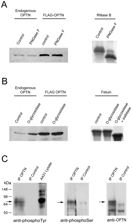
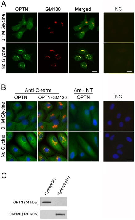
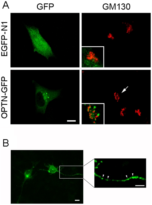
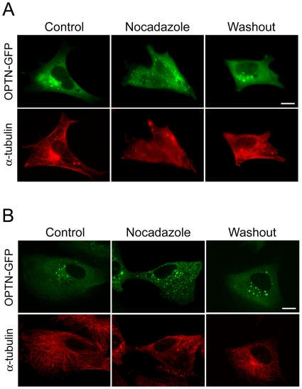
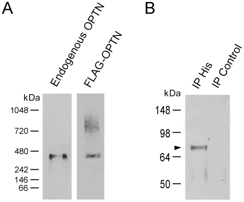
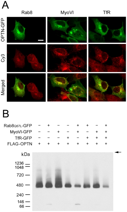
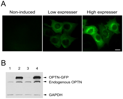
Methodology/principal findings: Lysates from rat retinal ganglion RGC5 cells were subjected to N- or O-deglycosylation or membrane protein extraction. The phosphorylation status was evaluated after immunoprecipitation. It was found that while phosphorylated, optineurin was neither N- nor O-glycosylated, and was by itself not a membrane protein. RGC5 and human retinal pigment epithelial cells were double stained with anti-optineurin and anti-GM130. The endogenous optineurin exhibited a diffuse, cytoplasmic distribution, but a population of the protein was associated with the Golgi apparatus. Turnover experiments showed that the endogenous optineurin was relatively short-lived, with a half-life of approximately 8 hours. Native blue gel electrophoresis revealed that the endogenous optineurin formed homohexamers. Optineurin also interacted with molecules including Rab8, myosin VI, and transferrin receptor to assemble into supermolecular complexes. When overexpressed, optineurin-green fluorescence protein (GFP) fusion protein formed punctate structures termed "foci" in the perinuclear region. Treatment of nocadazole resulted in dispersion of the optineurin foci. In addition, tetracycline-regulated optineurin-GFPs expressing RGC5 stable cell lines were established for the first time.

Conclusions/significance: The present study provides new information regarding basic characteristics of optineurin that are important for future efforts in defining precisely how optineurin functions normally and how mutations may result in pathology. The inducible optineurin-GFP-expressing cell lines are also anticipated to facilitate in-depth studies of optineurin. Furthermore, the demonstrations that optineurin is an aggregation-prone protein and that the foci formation is microtubule-dependent bear similarities to features documented in neurodegenerative diseases, supporting a neurodegenerative paradigm for glaucoma.


Conflict of interest statement

Competing Interests: The authors have declared that no competing interests exist.

Figures

Figure 1
Figure 1. Optineurin is neither N- (A) nor O- (B) glycosylated but is phosphorylated (C).
Lysates from RGC5 cells or those transfected with pOPTN-FLAG were untreated (control), or treated with PNGase F (for N-deglycosylation, A) or O-glycosidase (for O-deglycosylation) without (B) or with sialidase (not shown). The digested samples were subjected to Western blotting using anti-optineurin. The band pattern of optineurin (A and B, left panel) was not affected by any of the enzyme treatments. By Coomassie blue staining, the purified RNase B, as a positive control, had a band shift from 17 to 15 kDa after N-deglycosylation (A, right panel). The purified fetuin had a band shift from 64 to 55 kDa (B, right panel) after O-glycosidase and sialidase digestion. Fetuin was resistant to a single O-glycosidase digestion due to the fact that its N-linked and O-linked oligosaccharides are sialylated. C. Lysates from RGC5 cells were immunoprecipitated with either rabbit anti-C-terminal optineurin polyclonal antibody (IP OPTN) or normal rabbit IgG as an IP negative control (IP Control)) and were then immunoblotted with mouse anti-phosphotyrosine (anti-phosphoTyr), anti-mouse phosphoserine (anti-phosphoSer), or anti-C-terminal optineurin (anti-OPTN). The anti-optineurin but not rabbit IgG immunoprecipitated proteins were immunoreactive toward anti-phosphotyrosine (left panel) and anti-phosphoserine (middle panel). No immunoreactivity was observed with anti-phosphothreonine (not shown). Lysate from A431 (human epithelial carcinoma) cell line was used a positive control in anti-phosphotyrosine experiments. Immunoprecipitated pull down was verified by immunoblotting with anti-optineurin (anti-OPTN, right panel). Size markers are indicated. Arrows denote the 74-kDa optineurin band.
Figure 2
Figure 2. The endogenous optineurin is localized partially to the Golgi apparatus but is not a membrane protein.
A. RGC5 cells were double immunostained with anti-C-terminal optineurin (in green) and anti-GM130 (Golgi marker, in red). The endogenous optineurin (OPTN) had a diffuse cytoplasmic distribution pattern when 0.1 M glycine was included in the PBS rinsing buffer (top, left panel). Without glycine (bottom, left panel), the optineurin staining was prominent in the perinuclear region, colocalizing (in yellow, Merged) with the Golgi apparatus. Cells incubated with only secondary antibodies as negative controls (NC) showed no staining (right panels). B. Human RPE cells were immunostained with anti-C-terminal (Anti-C-term, in green) or anti-INT (Anti-INT, in green) optineurin along with anti-GM130 (in red). The endogenous optineurin (OPTN) had a diffuse cytoplasmic distribution pattern when anti-INT optineurin antibody was used (middle right panels) regardless whether glycine was included in the initial rinse. Using anti-C-terminal antibody, the optineurin staining was more perinuclar and Golgi-associated without the glycine rinse (left panels). Cells incubated with only secondary antibodies as negative controls (NC) showed minimal staining (right panels). The nuclei were stained with DAPI in blue. Scale bar, 10 µm. C. Western blot analyses of hydrophilic and hydrophobic fractions from RGC5 cell lysates. Total proteins in RGC lysates were subjected to membrane protein extraction. The hydrophilic fraction (left lane) contained predominantly non-membrane cytosolic proteins and the hydrophobic fraction (right lane) contained membrane proteins. Optineurin (OPTN) was detected exclusively in the hydrophilic fraction. GM130, a Golgi marker used as membrane protein positive control, was detected in the hydrophobic fraction as expected.
Figure 3
Figure 3. Cellular localization of overexpressed wild type optineurin.
A. RGC5 cells were transfected for 20 h, fixed and immunostained. When transfected with pEGFP-N1 (mock control, top, left panel), green fluorescence was observed in the entire RGC5 cells including the nucleus. When transfected with pOPTN-EGFP (OPTN-GFP, bottom, left panel), diffuse green fluorescence was likewise seen in the cytoplasm of RGC5 cells. In addition, bright granular or punctuate structures termed foci were also noted in the perinuclear region around the Golgi apparatus (stained with anti-GM130 in red). Insets show the enlarged and merged images in the perinuclear region. Optineurin foci largely did not overlap with the GM130 staining. Golgi fragmentation in pOPTN-EGFP-transfected cells (arrows) was observed. Scale bar, 10 µm. B. RGC5 cells were transfected with pOPTN-EGFP for 20 h and underwent differentiation with treatment of 316 nM staurosporine for 4 h. Punctate optineurin-GFP foci (green) were found on the neurite extensions (arrowheads). The enlarged and amplified image of the blocked area is shown in the right panel. Scale bars, 10 µm.
Figure 4
Figure 4. Distribution of optineurin foci is microtubule dependent.
RGC5 (A) and RPE (B) cells expressing optineurin-GFP (OPTN-GFP) were untreated (Control) or treated with 10 µM nocodazole for 30 min. The cells were washed thoroughly to remove nocodazole (Washout) and allowed to recover for 1 h. The optineurin-GFP foci appeared in green and the microtubule network was visualized by immunostaining with anti-α-tubulin in red. The microtubule was disrupted upon nocodazole treatment and the optineurin foci were dispersed from perinuclear region to distribute evenly in the cytoplasm. Upon nocadazole removal, the microtubule network was restored and the foci returned to the perinuclear area. Scale bar, 10 µm.
Figure 5
Figure 5. Oligomerization of optineurin.
A. Native blue gel electrophoresis. Total protein from RGC5 cells (30 µg) and that (15 µg) from RGC5 cells transfected for 20 h to express FLAG-optineurin (FLAG-OPTN) fusion protein were subjected to native blue gel electrophoresis and Western blotting. The membranes were probed either with anti-optineurin to detect the endogenous optineurin (left lane) or anti-FLAG to detect the forced expressed FLAG-OPTN fusion protein (right lane). A band at approximately 420 kDa was observed in both cases in the native gel. With the FLAG-OPTN, higher molecular weight bands which may represent the complex formed with other optineurin binding partners were additionally seen. B. Co-immunoprecipiation. RGC5 cells were co-transfected with pTarget-FLAG-OPTN and pOPTN-His. Cell lysates were immunoprecipitated with rabbit anti-His antibody, and the immunoprecipitated proteins were probed with anti-FLAG antibody under reducing conditions. An immuoreactive optineurin band (arrowhead) was detected in the anti-His (IP His, left lane) but not in the rabbit IgG (IP Control, right lane) pull down. Size markers are indicated.
Figure 6
Figure 6. Protein interactions of optineurin.
A. Colocalization of optineurin-GFP foci with Rab8, myosin VI (MyoVI) and transferrin receptor (TfR). RGC5 cells were transfected for 20 h to express optineurin-GFP (OPTN-GFP, in green) and immunostained with anti-Rab8, anti-MyoVI, or anti-TfR.Cy3-labled secondary antibody was used and the Rab8, MyoVI and TfR staining appeared in red (Cy3). Merged images are presented at the bottom panel. Colocalization of optineurin foci with Rab8, MyoVI, and TfR was observed in the perinuclear region in yellow in transfected cells. Those cells that stained positively with anti-Rab8, anti-MyoVI, and anti-TfR in red, but did not display any green fluorescence are non-transfected cells. B. Complex formation of optineurin with Rab8, MyoVI and TfR. RGC5 cells were co-transfected with p-Target-FLAG-OPTN (FLAG-OPTN) and pRab8Q67L-EGFP (Rab8Q67L-GFP), pMyoVI-EGFP (MyoVI-GFP), pTfR-EGFP (TfR-GFP), pRab8Q67L-EGFP + pMyoVI-EGFP, pRab8Q67L-EGFP + pTfR-EGFP, pMyoVI-EGFP+ pTfR-EGFP, or pRab8Q67L-EGFP + pMyoVI-EGFP + pTfR-EGFP. 20 µg of lysates from the transfected cells were subjected to native blue gel electrophoresis and Western blotting using anti-FLAG antibody. Super complexes with molecular sizes larger than 400 kDa were detected. The strongest complex formation was observed between FLAG-OPTN and MyoVI. The complex formation between them was reduced when Rab8Q67L and/or TfR was present. They may compete with each other to interact with optineurin. Size markers are indicated. Arrow denotes the sample loading front.
Figure 7
Figure 7. Inducible expression of optineurin-GFP in Tet-on RGC5 stable cell lines.
A. Expression of optineurin-GFP by fluorescent microscope in two clones of the stable cell lines. Upon Dox induction, the low expresser (middle panel) showed a faint level of fluorescence in the cytoplasm while the high expresser (right panel) displayed bright fluorescence with foci observed in the perinuclear region. Non-induced control with background level of optineurin-GFP is presented on the left panel. B. Western blotting confirmed the expression of optineurin-GFP (OPTN-GFP) fusion protein in the low (lane 2) and the high (lane 4) expressers upon Dox induction. The optineurin-GFP band is barely visible without Dox induction (lane 1 for low expresser and lane 3 for high expresser). The endogenous optineurin (endogenous OPTN) band was seen in all 4 lanes. Glyceraldehyde 3-phosphate dehydrogenase (GAPDH) was used as a loading control.
Figure 8
Figure 8. Turnover of endogenous and induced optineurin.
A. Turnover of the endogenous optineurin in RGC5 cells. The cells were treated with cycloheximide (5 µg/ml) for 4 h to block the protein synthesis, and were harvested at the indicated time points. Western blotting for optineurin (OPTN) and glyceraldehyde 3-phosphate dehydrogenase (GAPDH, for loading control) was performed and densitometry was carried out. The ratios between the level of optineurin and that of GAPDH are presented. The half life of the endogenous optineurin was determined to be approximately 8 h. B. Turnover of optineurin-GFP (OPTN-GFP) in low expressers. C. Turnover of optineurin-GFP (OPTN-GFP) in high expressers. The cells were treated for 20 h with Dox (1 µg/ml) to express optineurin-GFP. After exposing to cyclohexmide for 4 h, the lysates were collected at the indicated time points. Anti-optineurin was used to detect both OPTN-GFP and the endogenous optineurin (OPTN). Compared to RGC5 endogenous optineurin (A), the induced optineurin-GFP had a delayed degradation rate in both low and high expressers. The half life of optineurin-GFP was approximately 20 h in low expressers. It was prolonged to >24 h in high expressers.

References

    1. Allingham RR, Liu Y, Rhee DJ. The genetics of primary open-angle glaucoma: a review. Exp Eye Res. 2009;88:837–844. - PMC - PubMed
    1. Wiggs JL. Genetic etiologies of glaucoma. Arch Ophthalmol. 2007;125:30–37. - PubMed
    1. Fan BJ, Wang DY, Lam DS, Pang CP. Gene mapping for primary open angle glaucoma. Clin Biochem. 2006;39:249–258. - PubMed
    1. Wang N, Chintala SK, Fini ME, Schuman JS. Activation of a tissue-specific stress response in the aqueous outflow pathway of the eye defines the glaucoma disease phenotype. Nat Med. 2001;7:304–309. - PMC - PubMed
    1. Rezaie T, Child A, Hitchings R, Brice G, Miller L, et al. Adult-onset primary open-angle glaucoma caused by mutations in optineurin. Science. 2002;295:1077–1079. - PubMed

Publication types

MeSH terms

LinkOut - more resources