. 2010 Feb 11;5(2):e9159.
doi: 10.1371/journal.pone.0009159.

Ghrelin stimulation of growth hormone-releasing hormone neurons is direct in the arcuate nucleus

Affiliations

Ghrelin stimulation of growth hormone-releasing hormone neurons is direct in the arcuate nucleus

Guillaume Osterstock et al. PLoS One. .

Abstract

Background: Ghrelin targets the arcuate nucleus, from where growth hormone releasing hormone (GHRH) neurones trigger GH secretion. This hypothalamic nucleus also contains neuropeptide Y (NPY) neurons which play a master role in the effect of ghrelin on feeding. Interestingly, connections between NPY and GHRH neurons have been reported, leading to the hypothesis that the GH axis and the feeding circuits might be co-regulated by ghrelin.

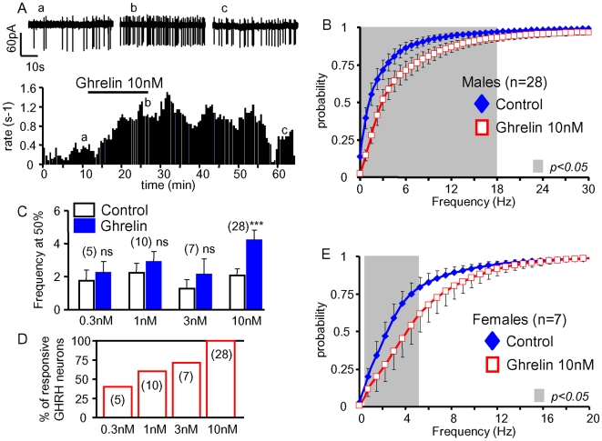
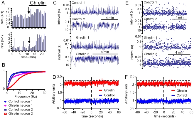
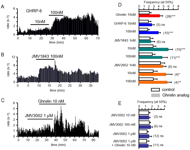
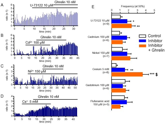
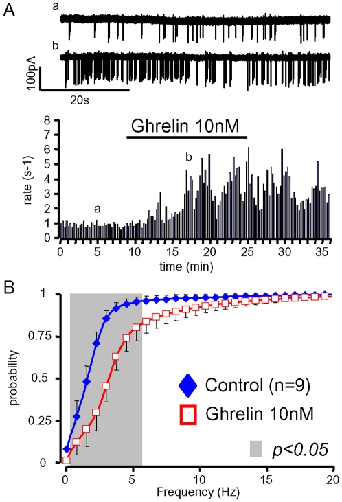
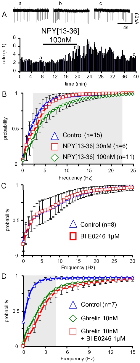
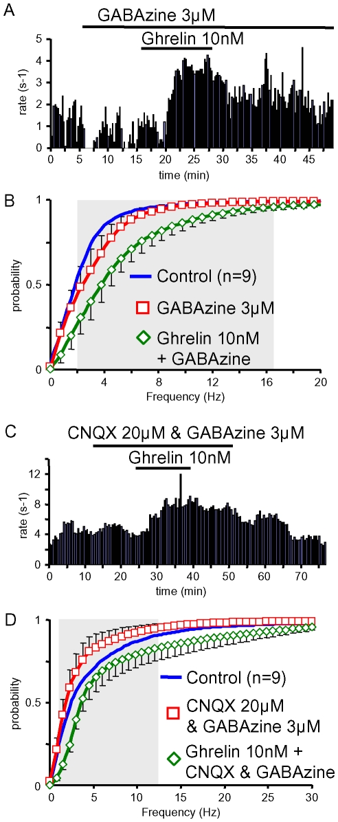
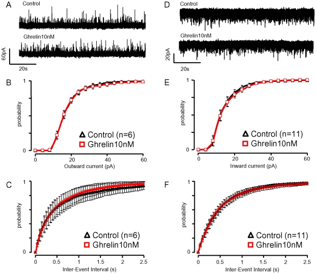
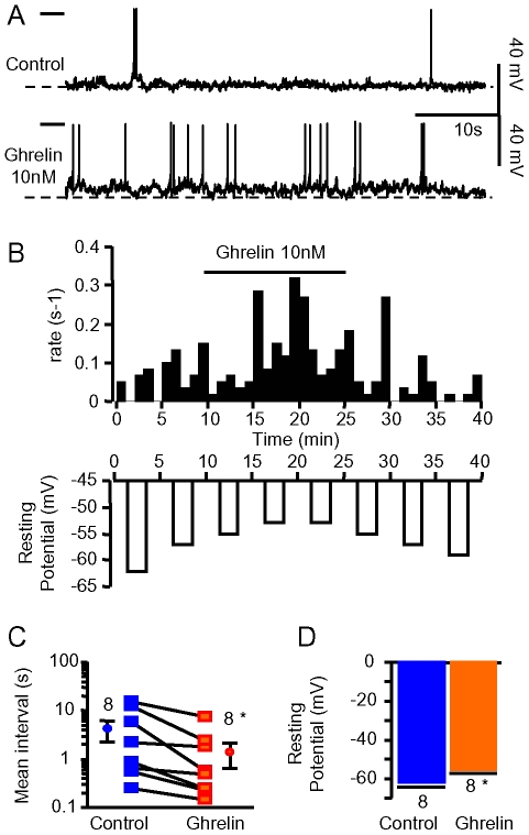
Principal findings: Here, we show that ghrelin stimulates the firing rate of identified GHRH neurons, in transgenic GHRH-GFP mice. This stimulation is prevented by growth hormone secretagogue receptor-1 antagonism as well as by U-73122, a phospholipase C inhibitor and by calcium channels blockers. The effect of ghrelin does not require synaptic transmission, as it is not antagonized by gamma-aminobutyric acid, glutamate and NPY receptor antagonists. In addition, this hypothalamic effect of ghrelin is independent of somatostatin, the inhibitor of the GH axis, since it is also found in somatostatin knockout mice. Indeed, ghrelin does not modify synaptic currents of GHRH neurons. However, ghrelin exerts a strong and direct depolarizing effect on GHRH neurons, which supports their increased firing rate.

Conclusion: Thus, GHRH neurons are a specific target for ghrelin within the brain, and not activated secondary to altered activity in feeding circuits. These results support the view that ghrelin related therapeutic approaches could be directed separately towards GH deficiency or feeding disorders.


Conflict of interest statement

Competing Interests: The authors have declared that no competing interests exist.

Figures

Figure 1
Figure 1. Ghrelin enhanced the activity of GHRH neurons.
A, time course of an experiment where the superfusion of a sagittal brain slice with 10 nM ghrelin increased, in a reversible manner, the rate of spontaneous action potentials of a GHRH neuron (individual traces shown on the top). B, summary of the effects of ghrelin (10 nM) on the cumulative distributions of action potential frequencies in GHRH neurons from adult males; C, mean effects of 0.3 to 10 nM ghrelin on the rate of spontaneous action potentials in GHRH neurons: the action potential frequencies observed at the half maximal values of the cumulated histograms were collected in each experiment in the absence and presence of ghrelin (see Methods for details). D, the proportion of stimulatory effects induced by ghrelin increased in a dose-dependent manner in GHRH neurons. E, summary of the effects of ghrelin (10 nM) on the distributions of action potential frequencies in GHRH neurons from adult females. In B & E, the symbols and lines are the means and sem. Statistical differences (p<0.05, paired student-t test) between curves are framed by the grey areas. In D, the bars and lines are the means and sem of the numbers of experiments indicated. ***, statistical difference from control values (p<0.001, paired student-t test).
Figure 2
Figure 2. Ghrelin did not synchronize the activity of GHRH neurons in dual patch-clamp epxeriments.
A, stimulatory effects of ghrelin (10 nM) on the firing rate of two GHRH neurons recorded simultaneously. Action potential rates were calculated every 30 s. B, cumulative distributions of the frequency of the action potentials of the GHRH neurons from panel A, showing the extent of the rightward shifts induced by ghrelin. C, intervals between action potentials of the GHRH neurons from panel A, under control conditions and in the presence of ghrelin, were then used in generating the cross-correlograms shown in D. The correlations of activity were calculated within consecutive bins of 100 ms during 60 s (see Methods for further details). Dotted lines indicate the 95% confidence boundaries within which the distributions behave as random, in the absence and presence of ghrelin. E&F, same as C&D, except that random distributions of instantaneous frequencies of action potentials were generated using the properties of the experimental data, in the absence and in the presence of ghrelin. The shapes of these cross-correlograms characterizing de-correlated series of events were almost undistinguishable from the experimental curves.
Figure 3
Figure 3. Effects of ghrelin receptor ligands on the activity of GHRH neurons.
A to C, typical experiments where superfusions with the agonists GHRP-6 (10 & 100 nM, A) and JMV1843 (100 nM, B) increased action potentials rates in adult male GHRH neurons; the antagonist JMV3002 (1 µM) prevented from the stimulatory effect of ghrelin (10 nM, C). D–E, summaries of the effects of GHSR agonists (D: ghrelin, GHRP-6, JMV1843, JMV2952) or antagonist (E: JMV3002) on the cumulative distributions of the spontaneous action potentials of GHRH neurons. The action potential frequencies observed at the half maximal values of the cumulated histograms were averaged according to the absence (white bars) and presence of agonists and/or antagonist (coloured bars, see Methods for details). In D&E, bars and lines are the means and the sem of the numbers of experiments indicated. Statistical differences *, p<0.05; **, p<0.01; ***, p<.005 are shown (paired student-t test).
Figure 4
Figure 4. The effect of ghrelin on GHRH neurons requires phospholipase C and calcium channels.
A–D, typical recordings from GHRH neurons in the absence and presence of the phospholipase C inhibitor U-73122 (10 µM, A); the high voltage-activated calcium channel blocker Cd2+ (100 µM, B); the low voltage-activated calcium channel blocker Ni2+ (150 µM, C); the HCN channel blocker Cs+ (5 mM, D); and 10 nM ghrelin (A–D), as indicated by the lines. E, summary of the effects of cellular signalling inhibitors on the cumulated distributions of spontaneous action potentials in GHRH neurons. The action potential frequencies observed at the half maximal values of the cumulated histograms were averaged according to the absence (white bars) and presence of inhibitor (blue bars), and in the presence of inhibitor plus 10 nM ghrelin (orange bars, see Methods for details). Bars and lines are the means and the sem of the numbers of experiments indicated. Statistical differences (vs control values *: p<0.05; **: p<0.01; ***, p<0.005; and vs inhibitor level $, p<0.05, paired student-t test) are shown.
Figure 5
Figure 5. Ghrelin enhances the firing rates of GHRH neurons in the absence of somatostatin.
A, typical experiment where 10 nM ghrelin increased the firing rate of a GHRH neuron from an adult male somatostatin −/−, GHRH-GFP mouse (raw traces are shown on the top). B, summary of the effects of ghrelin (10 nM) on the distributions of action potential frequencies in GHRH neurons from adult male somatostatin −/−, GHRH-GFP mice. Symbols and lines are the means and the sem of the numbers of experiments indicated. Statistical significances (p<0.05, paired student-t test, see methods) between curves are framed by the grey area.
Figure 6
Figure 6. The effect of ghrelin on the firing rates of GHRH neurons did not require Y-2 receptors.
A, typical experiment where the Y-2 receptors agonist, NPY – 100 nM, increased, in a reversible manner, the spontaneous firing rate in a male GHRH neuron. Raw traces are shown on top of the panel. B, summaries of the effects of NPY – (30 & 100 nM) on the distributions of action potential frequencies in GHRH neurons from adult male GHRH-GFP mice. C–D, summaries of the effects of the Y-2 antagonist BIIE0246 alone (C) and of ghrelin in the absence or presence of BIIE0246 (D) on the distributions of action potential frequencies in GHRH neurons from adult male GHRH-GFP mice. Symbols and lines are the means and the sem of the numbers of experiments indicated. Statistical significance (p<0.05, paired student-t test) between curves (effect of ghrelin in the absence or presence of BIIE0246, D) are framed by the grey area.
Figure 7
Figure 7. Ghrelin enhanced the firing rate of GHRH neurons during GABAA receptor inhibition.
A&B, typical experiments where spontaneous action potentials of GHRH neurons were recorded as ghrelin 10 nM was applied in the continuing presence of 3 µM GABAzine, a GABAA receptor antagonist, alone (A) or together with 20 µM CNQX, a AMPA/kainate receptors antagonist (B). C&D, summaries of the stimulatory effect of ghrelin (10 nM) on the distributions of action potential frequencies in GHRH neurons in the presence of 3 µM GABAzine (C) or in the presence of 3 µM GABAzine + 20 µM CNQX (D). Symbols and lines are the means and the sem of the numbers of experiments indicated. Statistical significance (p<0.05, paired student-t test) between mean values recorded in the presence of inhibitors alone and in the presence of inhibitors plus ghrelin is framed by the grey area. Note that the mean control distributions are shown as lines and sem omitted, for clarity.
Figure 8
Figure 8. Ghrelin did not modify spontaneous synaptic currents of GHRH neurons.
A&D, raw traces of spontaneous glutamatergic (−30 mV) and GABAergic (−70 mV) synaptic currents recorded in the absence and presence of 10 nM ghrelin in GHRH neurons from adult male GHRH-GFP mice. The effects of ghrelin (10 nM) were summarized, on the amplitude (B&E) and intervals (C&F) of glutamatergic (B&C) and GABAergic (E&F) synaptic currents. The cumulative distributions are represented as symbols and lines, i.e. the means and the sem of the numbers of experiments indicated.
Figure 9
Figure 9. Ghrelin changed the excitability of GHRH neurons.
A, recordings from a GHRH neuron in the absence and presence of 10 nM ghrelin, in the perforated patch-clamp configuration. B, time course of the effect of ghrelin 10 nM on the firing rate (upper graph) and on the resting potential (lower graph) of the GHRH neuron shown in A. C, summary of the effects of ghrelin 10 nM on the mean action potential intervals in GHRH neurons recorded in the perforated patch-clamp configuration. D, mean amplitude of the resting potential in GHRH neurons in the absence and presence of 10 nM ghrelin (same experiments as in C). Bars and lines are the means and the sem of the numbers of experiments indicated. Statistical difference (p<0.05, paired student-t test) with the control level is indicated.

Similar articles

Cited by

References

    1. Farhy LS, Bowers CY, Veldhuis JD. Model-projected mechanistic bases for sex differences in growth hormone regulation in humans. Am J Physiol Regul Integr Comp Physiol. 2007;292:R1577–93. - PubMed
    1. Abizaid A, Horvath TL. Brain circuits regulating energy homeostasis. Regul Pept. 2008;149:3–10. - PMC - PubMed
    1. Bowers CY, Momany FA, Reynolds GA, Hong A. On the in vitro and in vivo activity of a new synthetic hexapeptide that acts on the pituitary to specifically release growth hormone. Endocrinology. 1984;114:1537–45. - PubMed
    1. Smith RG. Development of growth hormone secretagogues. Endocr Rev. 2005;26:346–360. - PubMed
    1. Nakazato M, Murakami N, Date Y, Kojima M, Matsuo M, et al. A role for ghrelin in the central regulation of feeding. Nature London. 2001;409:194–198. - PubMed

Publication types

MeSH terms