. 2010 Feb 5:2:28.
doi: 10.3389/neuro.02.028.2009. eCollection 2009.

Cellular transport and membrane dynamics of the glycine receptor

Affiliations

Cellular transport and membrane dynamics of the glycine receptor

Andrea Dumoulin et al. Front Mol Neurosci. .

Abstract

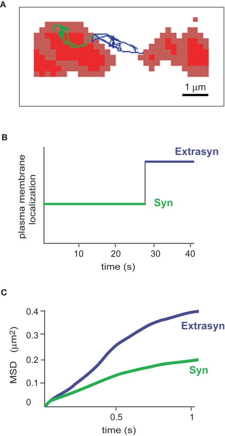
Regulation of synaptic transmission is essential to tune individual-to-network neuronal activity. One way to modulate synaptic strength is to regulate neurotransmitter receptor numbers at postsynaptic sites. This can be achieved either through plasma membrane insertion of receptors derived from intracellular vesicle pools, a process depending on active cytoskeleton transport, or through surface membrane removal via endocytosis. In parallel, lateral diffusion events along the plasma membrane allow the exchange of receptor molecules between synaptic and extrasynaptic compartments, contributing to synaptic strength regulation. In recent years, results obtained from several groups studying glycine receptor (GlyR) trafficking and dynamics shed light on the regulation of synaptic GlyR density. Here, we review (i) proteins and mechanisms involved in GlyR cytoskeletal transport, (ii) the diffusion dynamics of GlyR and of its scaffolding protein gephyrin that control receptor numbers, and its relationship with synaptic plasticity, and (iii) adaptative changes in GlyR diffusion in response to global activity modifications, as a homeostatic mechanism.

Keywords: SPT; cytoskeleton; diffusion; gephyrin; glycine receptor; motor proteins; neuronal activity; transport.


Figures

Figure 1
Figure 1
Gephyrin domains and structural organization. (A) Schematic depiction of the three gephyrin domains (G, C, E): the N-terminal G-domain (G) and the -terminal E-domain (E) are separated by a central C-domain (C). The C-domain is magnified below. Sequences of the binding sites for Pin1 (Zita et al., 2007), Dlc1/2 (Fuhrmann et al., 2002) and collybistin (Kins et al., 2000) are depicted by arrows. Numbers represent amino acid positions within the gephyrin protein. (B) The gephyrin «hexagonal lattice» oligomerization model: E-and G-gephyrin domains are able to dimerize and trimerize, respectively (left panel). Combination of these two properties would lead to a hexagonal structure of gephyrin (right panel) underneath the postsynaptic membrane, where GlyR (in black) would anchor itself through the binding of the intracellular loop of the beta subunit with the E-domain of gephyrin.
Figure 2
Figure 2
GlyR-gephyrin intracellular cytoskeleton transport. Newly synthesized glycine receptors (GlyRs) that leave the Golgi compartment reach the plasma membrane through active transport mechanisms along cytoskeletal elements. KIF5 motor proteins connect to vesicular GlyRs via gephyrin (green) that serves as a cargo adaptor in the transport complex. The KIF5/gephyrin/GlyR complex moves in anterograde directions toward the plus-ends of microtubules. It is currently unclear whether myosins mediate the final steps of GlyR surface membrane delivery and the initial steps of plasma membrane internalization, respectively, to traverse the submembrane actin cortex. At postsynaptic sites, gephyrin (green) forms a submembrane scaffold and mediates GlyR clustering. Exo-/ and endocytosis of receptors is thought to occur at extrasynaptic sites. Upon GlyR internalization, a GlyR/gephyrin/dynein transport complex mediates retrograde minus end-directed microtubule transport to intracellular compartments. Cytoplasmic dyneins are thought to participate in endocytic processes downstream on the sorting endosome (e.g. delivery to multivesicular bodies and/or lysososmes). In analogy to the anterograde GlyR transport complex, gephyrin (green) serves as a cargo adaptor that connects the vesicular receptor with its motor.
Figure 3
Figure 3
Activity-dependent polyglutamylation of tubulin alters intracellular transport. (A) Model of microtubule track changes through polyglutamylation (diagonal lines) and MAP2 binding (dark squares) upon altered neuronal activity. Increased activity, as induced through GlyR blockade (strychnine) or AMPAR activation (AMPA), interferes with gephyrin delivery into distal neurites (left). This effect is not observed upon neuronal activity reduction through AMPAR blockade (6,7-Dinitroquinoxaline-2,3-dione, DNQX) and can be prevented through functional depletion of neuronal polyglutamylase (the respective enzyme that adds polyglutamyl side chains to tubulin). Although it is unclear which modification is dominant, both represent negative signals for cargo delivery. (B) The individual cargo adaptor in the motor-cargo complex (gephyrin) is thought to mediate specificity of transport, as individual motor proteins transport multiple cargoes. Notably, KIF5-mediated transport of gephyrin is significantly reduced under strychnine conditions, whereas KIF5-mediated transport of GRIP1 (another cargo adaptor driven by the same motor) remains unaltered. The individual cargo adaptor within the transport complex (gephyrin) is therefore a candidate factor to sense modifications at the microtubule track surface (double arrow, question mark). Modified after Maas et al. (2009).
Figure 4
Figure 4
Diffusion properties of the glycine receptor. (A) Example of an individual GlyR-QDot trajectory exchanging between a synaptic (trace in green) and an extrasynaptic location (trace in blue). FM4-64-stained synapses are in red. (B) Time spent by the GlyR-QDot in the different compartments over a 40-s recording (same colour code). (C) Time-averaged MSD function of the QDot shown in (A). The two curves represent synaptic (green) and extrasynaptic (blue) portions of the trajectory. Curves are typical of confined (negatively bent) and free-diffusing molecules, respectively.
Figure 5
Figure 5
Model of receptor diffusion and stabilization at synapses. (A) Receptor exchanges between extrasynaptic and synaptic domains. The rates of entry and exit from gephyrin clusters define the kon and koff, respectively. (B) Representation of the different paths leading to the stabilization of GlyR by gephyrin clusters. Association of receptor (R) and its scaffolding protein gephyrin (S) can occur outside (equilibrium 1) or inside (equilibrium 4) synaptic sites. Once within clusters, receptor-scaffold complexes may reach a higher level of stabilization (equilibrium 5, dark gray). The index “i” indicates the inside and the index “o” the outside of the synaptic domain (light gray area) (modified from Ehrensperger et al., 2007).

References

    1. Adesnik H., Nicoll R. A., England P. M. (2005). Photoinactivation of native AMPA receptors reveals their real-time trafficking. Neuron 48, 977–985 10.1016/j.neuron.2005.11.030 - DOI - PubMed
    1. Allison D. W., Chervin A. S., Gelfand V. I., Craig A. M. (2000). Postsynaptic scaffolds of excitatory and inhibitory synapses in hippocampal neurons: maintenance of core components independent of actin filaments and microtubules. J. Neurosci. 20, 4545–4554 - PMC - PubMed
    1. Baas P. W., Deitch J. S., Black M. M., Banker G. A. (1988). Polarity orientation of microtubules in hippocampal neurons: uniformity in the axon and nonuniformity in the dendrite. Proc. Natl. Acad. Sci. U.S.A. 85, 8335–8339 10.1073/pnas.85.21.8335 - DOI - PMC - PubMed
    1. Bannai H., Lévi S., Schweizer C., Inoue T., Launey T., Racine V., Sibarita J. B., Mikoshiba K., Triller A. (2009). Activity-dependent tuning of inhibitory neurotransmission based on GABAAR diffusion dynamics. Neuron 62, 670–682 10.1016/j.neuron.2009.04.023 - DOI - PubMed
    1. Bats C., Groc L., Choquet D. (2007). The interaction between Stargazin and PSD-95 regulates AMPA receptor surface trafficking. Neuron 53, 719–734 10.1016/j.neuron.2007.01.030 - DOI - PubMed

LinkOut - more resources