

Further evidence for aberrant prefrontal salience coding in schizophrenia
- PMID: 20161811
- PMCID: PMC2821181
- DOI: 10.3389/neuro.08.062.2009
Further evidence for aberrant prefrontal salience coding in schizophrenia
Abstract
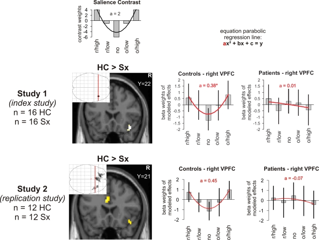
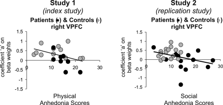
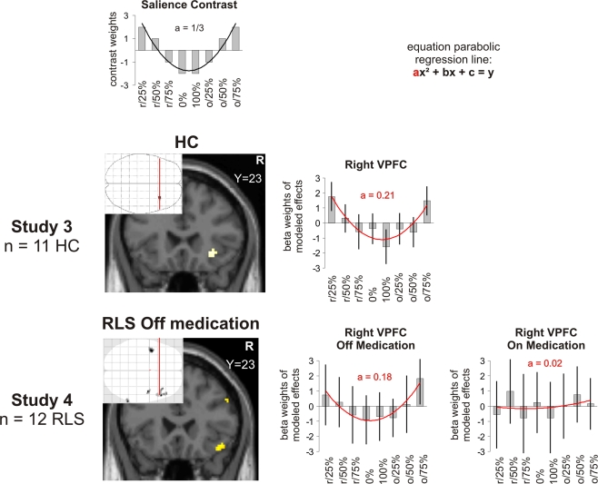
The revised dopamine hypothesis of schizophrenia postulates that dopamine metabolism is impacted differently with increased dopamine in the subcortical mesolimbic system and decreased dopamine in prefrontal cortical regions. Recently, we described findings supporting this hypothesis using a financial reward task in patients with schizophrenia (Walter et al., 2009). In addition to analysing prediction and prediction error coding, we found in this study evidence for aberrant cortical representation of salience in the right ventrolateral prefrontal cortex (VLPFC) in patients. Here, we reanalysed data of four other published reward studies of our group in order to investigate (i) whether we could replicate this finding in an independent cohort of patients with schizophrenia and (ii) how dopaminergic modulation impacts on cortical salience representation. Our main result was that we could replicate the finding of aberrant salience coding in the right VLPFC in patients with schizophrenia. Furthermore, we found evidence that the degree of salience coding in this region was correlated inversely with negative symptoms (anhedonia). Results of dopaminergic modulation showed tentative evidence for an influence of dopaminergic stimulation, but were not conclusive. In summary, we conclude that the right VLPFC might play a crucial role in salience coding and is impaired in schizophrenia.
Keywords: dopamine; functional magnetic resonance imaging; reward; salience; schizophrenia.
Figures
Similar articles
-
Dopaminergic dysfunction in schizophrenia: salience attribution revisited.Schizophr Bull. 2010 May;36(3):472-85. doi: 10.1093/schbul/sbq031. Epub 2010 May 7. Schizophr Bull. 2010. PMID: 20453041 Free PMC article. Review.
-
Altered reward functions in patients on atypical antipsychotic medication in line with the revised dopamine hypothesis of schizophrenia.Psychopharmacology (Berl). 2009 Sep;206(1):121-32. doi: 10.1007/s00213-009-1586-4. Epub 2009 Jun 12. Psychopharmacology (Berl). 2009. PMID: 19521678
-
Do patients with schizophrenia exhibit aberrant salience?Psychol Med. 2009 Feb;39(2):199-209. doi: 10.1017/S0033291708003863. Epub 2008 Jun 30. Psychol Med. 2009. PMID: 18588739 Free PMC article.
-
Expected value and prediction error abnormalities in depression and schizophrenia.Brain. 2011 Jun;134(Pt 6):1751-64. doi: 10.1093/brain/awr059. Epub 2011 Apr 10. Brain. 2011. PMID: 21482548
-
Dopaminergic basis of salience dysregulation in psychosis.Trends Neurosci. 2014 Feb;37(2):85-94. doi: 10.1016/j.tins.2013.11.003. Epub 2014 Jan 2. Trends Neurosci. 2014. PMID: 24388426 Review.
Cited by
-
Pavlovian reward prediction and receipt in schizophrenia: relationship to anhedonia.PLoS One. 2012;7(5):e35622. doi: 10.1371/journal.pone.0035622. Epub 2012 May 4. PLoS One. 2012. PMID: 22574121 Free PMC article.
-
Motivational Deficits in Schizophrenia and the Representation of Expected Value.Curr Top Behav Neurosci. 2016;27:375-410. doi: 10.1007/7854_2015_385. Curr Top Behav Neurosci. 2016. PMID: 26370946 Free PMC article. Review.
-
Hippocampal temporal-parietal junction interaction in the production of psychotic symptoms: a framework for understanding the schizophrenic syndrome.Front Hum Neurosci. 2012 Jun 22;6:180. doi: 10.3389/fnhum.2012.00180. eCollection 2012. Front Hum Neurosci. 2012. PMID: 22737114 Free PMC article.
-
Acute and Lifetime Stress and Psychotic Illness: The Roles of Reward and Salience Networks.J Psychiatr Brain Sci. 2022;7(6):e220012. doi: 10.20900/jpbs.20220012. Epub 2022 Dec 30. J Psychiatr Brain Sci. 2022. PMID: 36741029 Free PMC article.
-
The motivation and pleasure dimension of negative symptoms: neural substrates and behavioral outputs.Eur Neuropsychopharmacol. 2014 May;24(5):725-36. doi: 10.1016/j.euroneuro.2013.06.007. Epub 2014 Jan 22. Eur Neuropsychopharmacol. 2014. PMID: 24461724 Free PMC article. Review.
References
Grants and funding
LinkOut - more resources
Full Text Sources
Other Literature Sources