

A novel bioluminescent protease assay using engineered firefly luciferase
- PMID: 20161840
- PMCID: PMC2803436
- DOI: 10.2174/1875397300802010016
A novel bioluminescent protease assay using engineered firefly luciferase
Abstract
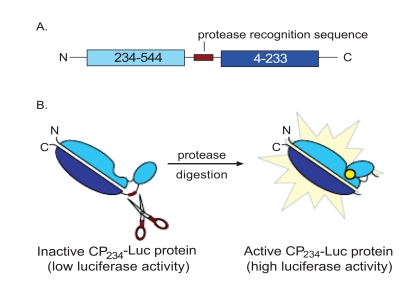
Proteases play important roles in a variety of disease processes. Understanding their biological functions underpins the efforts of drug discovery. We have developed a bioluminescent protease assay using a circularly permuted form of firefly luciferase, wherein the native enzyme termini were joined by a peptide containing a protease site of interest. Protease cleavage of these mutant luciferases greatly activates the enzyme, typically over 100 fold. The mutant luciferase substrates are easily generated by molecular cloning and cell-free translation reactions and thus the protease substrates do not need to be chemically synthesized or purchased. The assay has broad applicability using a variety of proteases and their cognate sites and can sensitively detect protease activity. In this report we further demonstrate its utility for the evaluation of protease recognition sequence specificity and subsequent establishment of an optimized assay for the identification and characterization of protease inhibitors using high throughput screening.
Figures


Similar articles
-
A bioluminescent assay for the sensitive detection of proteases.Biotechniques. 2011 Aug;51(2):105-10. doi: 10.2144/000113716. Biotechniques. 2011. PMID: 21806554
-
Engineering the C-terminus of firefly luciferase as an indicator of covalent modification of proteins.Biochim Biophys Acta. 1996 Jan 4;1292(1):89-98. doi: 10.1016/0167-4838(95)00199-9. Biochim Biophys Acta. 1996. PMID: 8547353
-
Bioluminescent assays for ADMET.Expert Opin Drug Metab Toxicol. 2008 Jan;4(1):103-20. doi: 10.1517/17425255.4.1.103. Expert Opin Drug Metab Toxicol. 2008. PMID: 18370862 Review.
-
Structural and functional effects of circular permutation on firefly luciferase: in vitro assay of caspase 3/7.Int J Biol Macromol. 2013 Jul;58:336-42. doi: 10.1016/j.ijbiomac.2013.04.015. Epub 2013 Apr 15. Int J Biol Macromol. 2013. PMID: 23597706
-
Building Biological Flashlights: Orthogonal Luciferases and Luciferins for in Vivo Imaging.Acc Chem Res. 2019 Nov 19;52(11):3039-3050. doi: 10.1021/acs.accounts.9b00391. Epub 2019 Oct 8. Acc Chem Res. 2019. PMID: 31593431 Free PMC article. Review.
Cited by
-
Tools for GPCR drug discovery.Acta Pharmacol Sin. 2012 Mar;33(3):372-84. doi: 10.1038/aps.2011.173. Epub 2012 Jan 23. Acta Pharmacol Sin. 2012. PMID: 22266728 Free PMC article. Review.
-
Imaging Proteolytic Activities in Mouse Models of Cancer.Methods Mol Biol. 2018;1731:247-260. doi: 10.1007/978-1-4939-7595-2_22. Methods Mol Biol. 2018. PMID: 29318559 Free PMC article.
-
Imaging proteolytic activity in live cells and animal models.PLoS One. 2013 Jun 11;8(6):e66248. doi: 10.1371/journal.pone.0066248. Print 2013. PLoS One. 2013. PMID: 23776643 Free PMC article.
-
Bioluminescence technologies to detect calicivirus protease activity in cell-free system and in infected cells.Antiviral Res. 2011 Apr;90(1):9-16. doi: 10.1016/j.antiviral.2011.02.002. Epub 2011 Feb 21. Antiviral Res. 2011. PMID: 21316392 Free PMC article.
-
Modular Peroxidase-Based Reporters for Detecting Protease Activity and Protein Interactions with Temporal Gating.J Am Chem Soc. 2022 Dec 21;144(50):22933-22940. doi: 10.1021/jacs.2c08280. Epub 2022 Dec 13. J Am Chem Soc. 2022. PMID: 36511757 Free PMC article.
References
-
- Hooper NM. Proteases in biology and medicine. London: Portland Press; 2002.
-
- Bogdanovic S, Langlands B. Westborough: D&MD Publications; 2005. Proteases: Technologies and opportunities for drug discovery.
-
- Timmer JC, Salvesen GS. Caspase substrates. Cell Death Differ. 2007;14:66–72. - PubMed
-
- Thormberry NA, Rano TA, Peterson EP, et al. A combinatorial approach defines specificities of members of the caspase family and granzyme B. J Biol Chem. 1997;272:17907–11. - PubMed
LinkOut - more resources
Full Text Sources
Other Literature Sources