

A review of the scientific basis and practical application of a new test of utricular function--ocular vestibular-evoked myogenic potentials to bone-conducted vibration
- PMID: 20161874
- PMCID: PMC2816364
A review of the scientific basis and practical application of a new test of utricular function--ocular vestibular-evoked myogenic potentials to bone-conducted vibration
Abstract
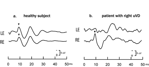
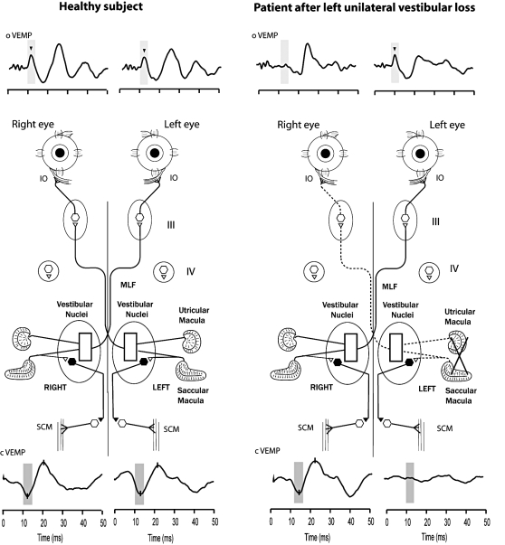
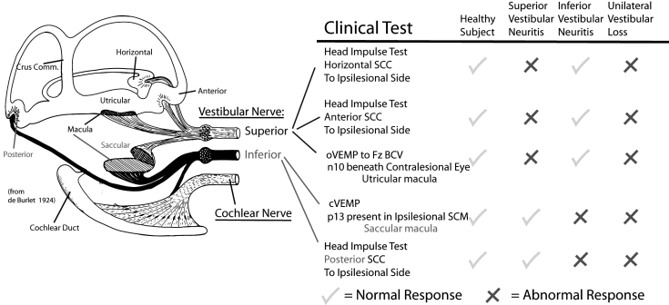
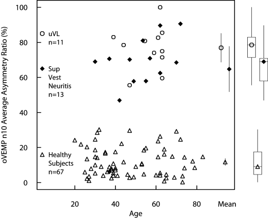
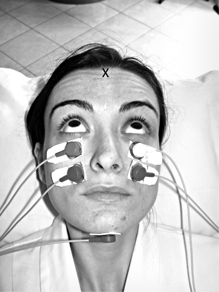
This is a review of recently published papers showing that bone-conducted vibration of the head causes linear acceleration stimulation of both inner ears and this linear acceleration is an effective way of selectively activating otolithic afferent neurons. This simple stimulus is used in a new test to evaluate clinically the function of the otoliths of the human inner ear. Single neuron studies in animals have shown that semicircular canal neurons are rarely activated by levels of bone-conducted vibration at 500 Hz which generate vigorous firing in otolithic irregular neurons and which result in a variety of vestibulo-spinal and vestibulo-ocular responses, and the latter is the focus of this review. In humans, 500 Hz bone-conducted vibration, delivered at the midline of the forehead, at the hairline (Fz), causes simultaneous and approximately equal amplitude linear acceleration stimulation at both mastoids and results in ocular-evoked myogenic potentials (oVEMPs) beneath both eyes. The first component of this myogenic potential, at a latency to peak of about 10 ms is a negative potential and is called n10 and, in healthy subjects, is equal in amplitude beneath both eyes, but after unilateral vestibular loss, the n10 potential beneath the eye opposite to the lesioned ear is greatly reduced or totally absent. n10 is a myogenic potential due to a crossed otolith-ocular pathway. In patients with total unilateral superior vestibular neuritis, in whom saccular function is largely intact (as shown by the presence of cervical vestibular evoked myogenic potentials (cVEMPs), but utricular function is probably compromised, there is a reduced n10 response beneath the contralesional eye, strongly indicating that n10 is due to utricular otolithic function.
La vibrazione portata al cranio (bone-conducted vibration) determina una stimolazione pari ad una accelerazione lineare di entrambi gli orecchi interni. Queste accelerazioni lineari sono in realtà un vero e proprio modo per attivare selettivamente i neuroni provenienti dalle macule utricolo sacculari. Lo studio di singoli neuroni nell’animale ha dimostrato che i neuroni dei canali semicircolari sono raramente attivati dai livelli di vibrazione che invece generano una vigorosa scarica nervosa da parte dei neuroni otolitici di tipo irregolare. In tal modo, l’attivazione otolitica indotta dalla vibrazione ossea risulta in una varietà di risposte di tipo vestibolo-spinale e vestibolo-oculare e proprio queste ultime, le vestibolo oculari, sono l’argomento di questa review. La vibrazione portata al cranio portata alla posizione della testa che coincide con la linea mediana in corrispondenza dell’inserzione-attaccatura dei capelli (Fz) causa un’accelerazione lineare simultanea ed approssimativamente uguale in ampiezza in corrispondenza di entrambe le mastoidi e risulta in potenziali evocati miogenici (oVEMPs) registrati sotto gli occhi nei soggetti sani. La prima componente di questo potenziale (n10) è uguale in ampiezza se registrata sotto gli occhi, ma dopo la perdita della funzione vestibolare l’onda potenziale n10 registrata sotto l’occhio opposto all’orecchio sede di lesione è grandemente e fortemente ridotta o del tutto assente. Questo risultato è dovuto all’esistenza di una via crociata otolito-oculare. Nei pazienti con esiti di nevrite del nervo vestibolare superiore nei quali la funzione sacculare è intatta, ma la funzione utricolare è compromessa, c’è una riduzione in ampiezza dell’onda n10 registrata sotto l’occhio opposto al lato leso, ciò sta fortemente a significare che l’onda potenziale n10 è dovuta alla funzione otolitica della macula utricolare.
Keywords: Bone conduction; Ocular vestibular-evoked myogenic potentials; Otolith; Vestibular disorder; Vestibular-evoked myogenic potentials.
Figures
References
-
- Halmagyi GM, Curthoys IS. A clinical sign of canal paresis. Arch Neurol 1988;45:737-9. - PubMed
-
- Halmagyi GM, Yavor RA, Colebatch JG. Tapping the head activates the vestibular system: a new use for the clinical reflex hammer. Neurology 1995;45:1927-9. - PubMed
-
- Rosengren SM, Todd NPM, Colebatch JG. Vestibular-evoked extraocular potentials produced by stimulation with bone-conducted sound. Clin Neurophysiol 2005;116:1938-48. - PubMed
-
- Todd NPM, Rosengren SM, Aw ST, Colebatch JG. Ocular vestibular evoked myogenic potentials (OVEMPs) produced by air- and bone-conducted sound. Clin Neurophysiol 2007;118:381-90. - PubMed
Publication types
MeSH terms
LinkOut - more resources
Full Text Sources