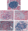
Protective effect of quercetin on the morphology of pancreatic beta-cells of streptozotocin-treated diabetic rats
- PMID: 20162074
- PMCID: PMC2816429
- DOI: 10.4314/ajtcam.v4i1.31196
Protective effect of quercetin on the morphology of pancreatic beta-cells of streptozotocin-treated diabetic rats
Abstract
This study was undertaken to investigate the protective effects of quercetin (QCT) on the morphology of pancreatic beta-cells against diabetes mellitus and oxidative stress experimentally-induced by streptozotocin (STZ) treatment in Wistar rats. Fifty male and female Wistar rats (200-250 g) were randomly divided into three experimental groups (i. e., control, STZ-treated, and STZ + Quercetin-treated groups). Diabetes was induced in the diabetic groups (B and C) of animals, by a single intraperitoneal injection of STZ (75 mg/kg), while each of the rats in the 'control' group received equal volume of citrate buffer (pH 6.3) solution intraperitoneally. In group C rats, quercetin (QCT, 25 mg/kg/day i.p.) was injected daily for 3 days prior to STZ treatment, and QCT administration continued until the end of the study period (30 days). Diabetes mellitus was confirmed by using Bayer's Glucometer Elite and compatible blood glucose test strips. The rats were sacrificed serially until the end of the study period (after 30 days). The pancreases of the sacrificed rats were excised and randomly processed for histological staining and biochemical assays for antioxidant enzymes [such as glutathione peroxidase (GSHPx), superoxide dismutase (SOD), catalase (CAT), malondialdehyde (MDA) and serum nitric oxide (NO)]. In the diabetic state, pancreatic beta-cells of STZ-treated group B rats histologically demonstrated an early chromatin aggregation, cytoplasmic vesiculation in the central beta-cells, nuclear shrinkage, and lysis of beta-cells with distortion of granules. The morphology of QCT-treated rats' pancreases showed viable cellularity with distinct beta-cell mass. STZ treatment significantly decreased (p<0.05) GSHPx, SOD, CAT and pancreatic insulin content. However, STZ treatment increased blood glucose concentrations, MDA and serum NO. The QCT-treated group of animals showed a significant decrease (p<0.05) in elevated blood glucose, MDA and NO. Furthermore, QCT treatment significantly increased (p<0.05) antioxidant enzymes' activities, as well as pancreatic insulin contents. Quercetin (QCT) treatment protected and preserved pancreatic beta-cell architecture and integrity. In conclusion, the findings of the present experimental animal study indicate that QCT treatment has beneficial effects on pancreatic tissues subjected to STZ-induced oxidative stress by directly quenching lipid peroxides and indirectly enhancing production of endogenous antioxidants.
Keywords: Antioxidant enzymes; Pancreatic β-cell; Quercetin; Streptozotocin.
Figures


References
-
- Bach J F. Insulin-dependent diabetes mellitus as an autoimmune disease. Endicr Rev. 1994;15:516–542. - PubMed
-
- Baynes J W. Role of oxidative stress in the development of complications in diabetes. Diabetes. 1991;40:405–412. - PubMed
-
- Bernard-Kargar C, Ktorza A. Endocrine pancreas plasticity under physiological and pathological conditions. Diabetes. 2001;50:30S–35S. - PubMed
-
- Bronner C, Landry Y. Kinetics of inhibitory effect of flavonoids on histamine secretion from mast cells. Agents Action. 1985;16:147–151. - PubMed
-
- Butler E A, Janson J, Bonner-Weir S, Ritzel R, Rizza R A, Butler P C. Beta-cell deficit and increased β-cell apoptosis in humans with type2 diabetes. Diabates. 2003;52:102–110. - PubMed
LinkOut - more resources
Full Text Sources
Miscellaneous
