. 2010 Jun;119(3):311-24.
doi: 10.1007/s00412-010-0258-9. Epub 2010 Feb 17.

Meiotic silencing and fragmentation of the male germline restricted chromosome in zebra finch

Affiliations

Meiotic silencing and fragmentation of the male germline restricted chromosome in zebra finch

Sam Schoenmakers et al. Chromosoma. 2010 Jun.

Abstract

During male meiotic prophase in mammals, X and Y are in a largely unsynapsed configuration, which is thought to trigger meiotic sex chromosome inactivation (MSCI). In avian species, females are ZW, and males ZZ. Although Z and W in chicken oocytes show complete, largely heterologous synapsis, they too undergo MSCI, albeit only transiently. The W chromosome is already inactive in early meiotic prophase, and inactive chromatin marks may spread on to the Z upon synapsis. Mammalian MSCI is considered as a specialised form of the general meiotic silencing mechanism, named meiotic silencing of unsynapsed chromatin (MSUC). Herein, we studied the avian form of MSUC, by analysing the behaviour of the peculiar germline restricted chromosome (GRC) that is present as a single copy in zebra finch spermatocytes. In the female germline, this chromosome is present in two copies, which normally synapse and recombine. In contrast, during male meiosis, the single GRC is always eliminated. We found that the GRC in the male germline is silenced from early leptotene onwards, similar to the W chromosome in avian oocytes. The GRC remains largely unsynapsed throughout meiotic prophase I, although patches of SYCP1 staining indicate that part of the GRC may self-synapse. In addition, the GRC is largely devoid of meiotic double strand breaks. We observed a lack of the inner centromere protein INCENP on the GRC and elimination of the GRC following metaphase I. Subsequently, the GRC forms a micronucleus in which the DNA is fragmented. We conclude that in contrast to MSUC in mammals, meiotic silencing of this single chromosome in the avian germline occurs prior to, and independent of DNA double strand breaks and chromosome pairing, hence we have named this phenomenon meiotic silencing prior to synapsis (MSPS).


Figures
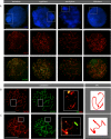
Fig. 1
Fig. 1
The GRC throughout meiotic prophase in zebra finch spermatocytes. a Overview of the different stages of meiotic prophase I of zebra finch spermatocytes. The top panel shows the DAPI staining of the different nuclei, the lower panel shows the corresponding nuclei stained for SYCP3 (red), the bottom panel shows the same nuclei stained for both SYCP3 (red) and γH2AX (green). The position of the GRC is indicated by a box. Bar represents 10 μm. b, c Spermatocyte spread nuclei immunostained for SYCP3 (red) and SYCP1 (green). The GRC is boxed. Enlargement and the schematic drawing of GRC are shown. No (b) or a small fragment (c) of SYCP1 signal is present on the GRC. Bar represents 5 μm
Fig. 2
Fig. 2
The GRC and meiotic recombination. a Spread nuclei immunostained for DAPI (blue), γH2AX (green) and SYCP3 (red). After metaphase I, the GRC becomes strongly positive for γH2AX. The GRC is boxed. Bar represents 5 μm. Sec spc secondary spermatocyte. b Spermatocyte spread nucleus immunostained for SYCP3 (red), RAD51 (green) and DAPI (blue). The GRC is boxed. Most RAD51 foci localise on axial elements of the SC. The bottom image represents a magnification of the area of the boxed GRC. Few small RAD51 foci are visible on the GRC. Bar represents 10 μm. c Spermatocyte spread nucleus immunostained for SYCP3 (red), MLH1 (green) and DAPI (blue). The area of the GRC is boxed. The bottom image represents a magnification of the area of the boxed GRC, and shows the presence of a MLH1 focus on the GRC. Bar represents 10 μm
Fig. 3
Fig. 3
The GRC is silenced during early meiotic prophase. ad Preleptotene spermatocyte spread nuclei. Bar represents 5 μm. a Top panel shows early and late preleptotene nuclei stained for SYCP3 (red) and DAPI (blue). The DAPI dense GRC localisation in late preleptotene stages is indicated with an asterisk; the lower panel shows only the DAPI staining. The early preleptotene nucleus shows no heterochromatic area, indicating that the DDB has not yet formed. The formation of a few SYCP3 axial element fragments in all nuclei marks the preleptotene stage. b The top panel shows a mid and late preleptotene nucleus stained for SYCP3 (red) and DAPI (blue). The asterisk indicates the DAPI dense GRC. The lower panel shows the same nuclei, but immunostained for γH2AX (green), indicative of the presence of the first DNA double strand breaks that more or less colocalise with SYCP3. c, d The top panel shows early and late preleptotene stages with DAPI staining. The asterisk indicates the DAPI dense GRC. The lower panel shows the corresponding nuclei stained for H3K9me3 (c) and macroH2A (d). Early preleptotene nuclei lack intense H3K9me3 (c) and macroH2A (d) staining, and no DDB is observed. e The top panel shows DAPI staining of the different nuclei, the middle panel shows the corresponding nuclei stained for H3K9me3 (red) and the bottom panel shows the merge. In preleptotene, a faint staining is present throughout the nucleus. In all consecutive meiotic prophase stadia, the GRC is positive for H3K9me3. Sec spc secondary spermatocyte. Bar represents 5 μm
Fig. 4
Fig. 4
The silenced GRC is eliminated at metaphase I in the vast majority of spermatocytes. a Spermatocyte spread nuclei stained for DAPI (blue, upper and lower panels) and SYCP3 (lower panel, red). The left image shows a secondary spermatocyte with the GRC still inside the nucleus, the middle image with the GRC still attached to the outside of the nucleus and the right image shows a secondary spermatocyte lacking the GRC. The typical SYCP3 filaments/dots mark the secondary spermatocyte stage. F the number of positive nuclei/the total number of scored secondary spermatocyte nuclei. Bar represents 5 μm. b, c, d Male meiotic prophase spread nuclei stained for H2AK119 ubiquitylation (green, b), sumoylation (green, c), H4K16ac (green, d) and SYCP3 (red). At pachytene, the GRC becomes increasingly sumoylated, and acetylated on H4K16. However, no H2AK119 ubiquitylation signal is present on the GRC at this stage. When the GRC is almost eliminated, it becomes increasingly ubiquitylated at H2AK119 (b). The area of the GRC is encircled. Bar represents 5 μm
Fig. 5
Fig. 5
The GRC is eliminated during the first meiotic division, and its DNA is degraded. a Male meiotic prophase spread nuclei stained for INCENP (green) and DAPI (blue). In pachytene, INCENP foci most likely follow the axial elements, which can be inferred from the DAPI staining. Also, many INCENP foci are present throughout the nucleus. The GRC remains largely devoid of INCENP staining. From diplotene onward, INCENP foci relocalise and concentrate mainly around the centromeres in metaphase. The GRC mostly shows no INCENP staining at its centromere. The area of the GRC is boxed. Bar represents 5 μm. b Spermatocyte spread nuclei stained for TUNEL (red) and DAPI (blue). Early in meiosis the GRC is largely devoid of TUNEL staining. When the GRC is almost expelled from the nucleus, it becomes strongly TUNEL-positive. The area of the GRC is boxed. Bar represents 5 μm
Fig. 6
Fig. 6
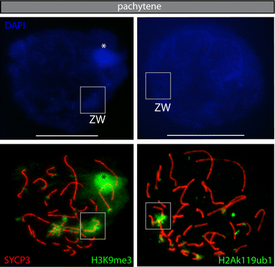
Analyses of the ZW pair and GRC chromosomes during female meiotic prophase. Oocyte spread nuclei stained for SYCP3 (red), H3K9me3 (green), H2AK119ub (green) and DAPI (blue). At pachytene, the ZW pair is strongly positive for H3K9me3 and H2Ak119ubiquitylation. Asterisk indicates the middle part of the GRC, which is also positive for H3K9me3, indicating that the GRC is also (partially) silenced during female meiotic prophase. GRC is boxed. Bar represents 5 μm
Fig. 7
Fig. 7
Schematic overview of the behaviour of the GRC during male meiosis of the zebra finch. In the transition phase between spermatogonia and the start of the male meiotic prophase, the GRC is identified and from preleptotene, it becomes heterochromatised and gains specific chromatin modifications such as H3K9me3, macroH2A, sumoylation and H4K16ac. With progression through meiotic prophase, all chromatin of the GRC is drawn into a heterochromatic body at the periphery of the nucleus. Few DSBs are induced on the GRC, and these remain unrepaired. Also, H2A on its centromere is not ubiquitylated. Sumoylation of chromatin and acetylation at H4K16 accumulate on the GRC and reach a maximum around mid pachytene. At diplotene, the GRC becomes acetylated at H3K9 and starts to decondense. During metaphase I, the nuclear membrane is degraded, the centromere and kinetochore of the GRC are aberrant and proper attachment to the spindles fails. This may cause DNA degradation of the GRC, and the chromatin to become phosphorylated, ubiquitylated and again de-ubiquitylated. In addition, the GRC forms a micronucleus with ongoing DNA fragmentation in the cytoplasm of the secondary spermatocyte

Similar articles

Cited by

References

    1. Anderson LK, Reeves A, Webb LM, Ashley T. Distribution of crossing over on mouse synaptonemal complexes using immunofluorescent localization of MLH1 protein. Genetics. 1999;151:1569–1579. - PMC - PubMed
    1. Ashley T, Plug AW, Xu J, Solari AJ, Reddy G, Golub EI, Ward DC. Dynamic changes in Rad51 distribution on chromatin during meiosis in male and female vertebrates. Chromosoma. 1995;104:19–28. doi: 10.1007/BF00352222. - DOI - PubMed
    1. Baker SM, Plug AW, Prolla TA, Bronner CE, Harris AC, et al. Involvement of mouse Mlh1 in DNA mismatch repair and meiotic crossing over. Nat Genet. 1996;13:336–342. doi: 10.1038/ng0796-336. - DOI - PubMed
    1. Bernstein E, Allis CD. RNA meets chromatin. Genes Dev. 2005;19:1635–1655. doi: 10.1101/gad.1324305. - DOI - PubMed
    1. Cabrero J, Teruel M, Carmona FD, Jimenez R, Camacho JP. Histone H3 lysine 9 acetylation pattern suggests that X and B chromosomes are silenced during entire male meiosis in a grasshopper. Cytogenet Genome Res. 2007;119:135–142. doi: 10.1159/000109630. - DOI - PubMed

Publication types