Classical Article
. 2010 Apr;468(4):1056-65.
doi: 10.1007/s11999-010-1239-2.

The classic: on the inner architecture of bones and its importance for bone growth. 1870

Classical Article

The classic: on the inner architecture of bones and its importance for bone growth. 1870

Julius Wolff. Clin Orthop Relat Res. 2010 Apr.

Abstract

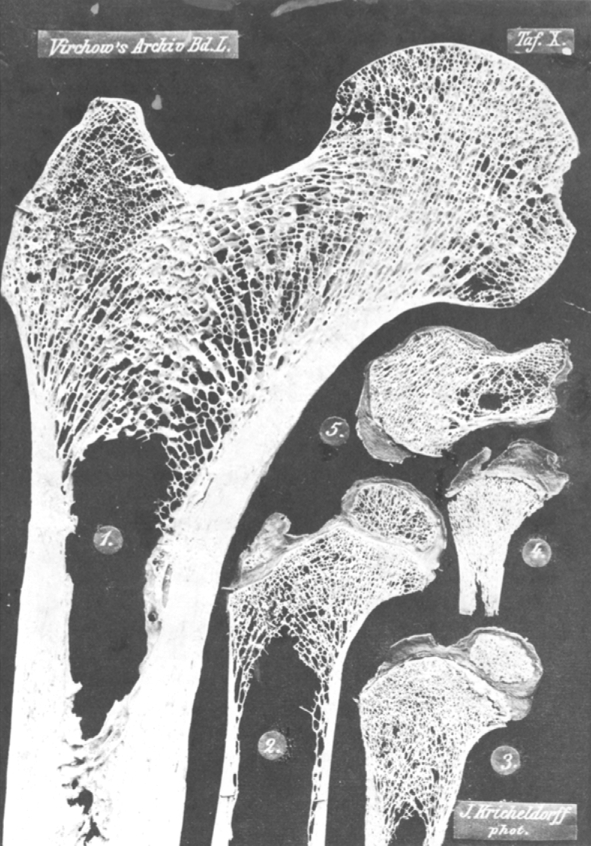
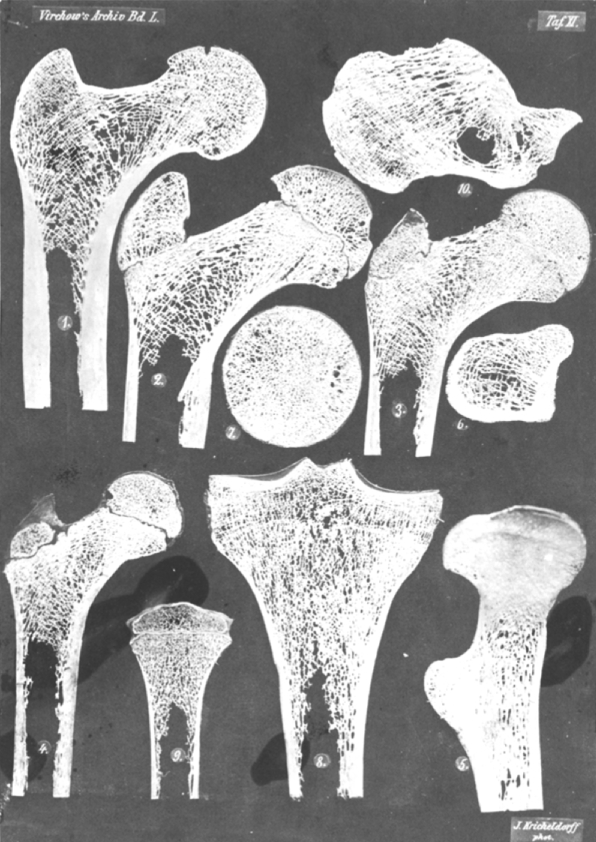
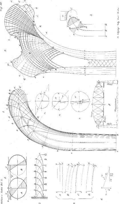
This Classic article is a translation and abridgment by M.O. Heller, W.R. Taylor, N. Aslanidis, and Georg N. Duda of the original work by Julius Wolff, Ueber die Innere Architectur der Knochen und ihre Bedeutung für die Frage vom Knochenwachstum (supplemental materials are available with the online version of CORR). An accompanying biographical sketch on Julius Wolff is available at DOI 10.1007/s11999-010-1258-z. A second Classic article is available at DOI 10.1007/s11999-010-1240-9. An accompanying Editorial is available at DOI 10.1007/s11999-010-1238-3. The Classic Article is ©1870 and is reprinted from Wolff J. Ueber die innere Architectur der Knochen und ihre Bedeutung für die Frage vom Knochenwachsthum. Virchows Arch Pathol Anat Physiol. 1870;50:389–450.

Electronic supplementary material: The online version of this article (doi:10.1007/s11999-010-1239-2) contains supplementary material, which is available to authorized users.


Figures

Table X
Table X
The images in this table have been photographed in natural size. The specimens depicted in Figures 1, 2 and 5 have been cut on the ivory cutting machine, those in Figures 3 and 4 have been cut freehand. Figure 1. Frontal longitudinal thin section, somewhat eccentric, closer to the dorsal surface, from the upper end of the right proximal femur of a 31-year-old man. Figure 2. A similar, but more centrally set longitudinal section from the proximal femur of a 3-year-old girl. Figure 3. The same from a 1¼-year-old boy. Figure 4. The same from a newborn boy. Figure 5. Sagittal longitudinal section through the middle of the Calcaneus of a 5-year-old girl.
Table XI
Table XI
All images in this table have been photographed in exactly half their natural size. It is advantageous to regard this table through a magnifying glass, for everything that might have been lost through the reduced depiction is completely recreated by this method of observation. The specimens depicted in Figures 1–4 and 8–10 have been cut on the ivory cutting machine, that in Figure 5 on the mechanical fret saw and those in Figures 6 and 7 by freehand. Figure 1. Frontal central longitudinal section from the femur of a 25-year-old woman. Figure 2. The same from the femur of a 20-year-old man. Figure 3. The same from a 13-year-old boy. Figure 4. The same from a 7-year-old girl. Figure 5. Sagittal longitudinal section, following the bent axis of the bone and thus depicting its neutral fiber layer, from the left proximal femur of a 29 year old man, seen from the concave (the medial, compression or adductor) side. The side of the specimen which intersects the dorsal surface of the proximal femur with the trochanter minor lies to the left. Figure 6. Cross-section (at a right angle to the axis) of the right proximal femur of the same man, at the height of the trochanter minor, seen from above. The side of the specimen which corresponds to the frontal surface of the proximal femur lies towards the lower side of the page, that of the dorsal surface (with the trochanter minor) towards the top, the tensile side respectively on the left, the compression side on the right. Figure 7. Cross-section of the same proximal femur, out of the femoral head, roughly in the region between its middle and lower third, seen from below. Here the side corresponding to the frontal surface of the proximal femur is again at the bottom, the side corresponding to the dorsal surface at the top, the tensile side at the left and the compressive side at the right. Figure 8. Frontal longitudinal section through the upper end of the lower leg of a 28-year-old woman. Figure 9. The same from a 5-year-old girl. Figure 10. Sagittal longitudinal section through the middle of the calcaneus of a 35-year-old man.
Table XII
Table XII
Figure 1. Depiction of the forces and trajectories in effect inside a bone. Based on the original drawn by the students of professor Culmann and under his supervision in double the original size of a human proximal femur. This original drawing was first photographically reduced back to the natural size and later lithographed. Figures 1a, b and c depict the force distribution for the exemplary chosen cross sections I, III and VI. Figure 2. Schematic reproduction of the specimen photographed in Figure 1, Table X. Figures 3–7 are part of the explanations on graphical statics on pp. 402–407. Figure 8. Schematic depiction of a bridge built according to Pauly’s system.

Similar articles

Cited by

Personal name as subject