. 2010 Jun;40(3):307-19.
doi: 10.1007/s11262-010-0455-x. Epub 2010 Feb 17.

Characteristics of Epstein-Barr virus envelope protein gp42

Affiliations

Characteristics of Epstein-Barr virus envelope protein gp42

Pamela L Shaw et al. Virus Genes. 2010 Jun.

Abstract

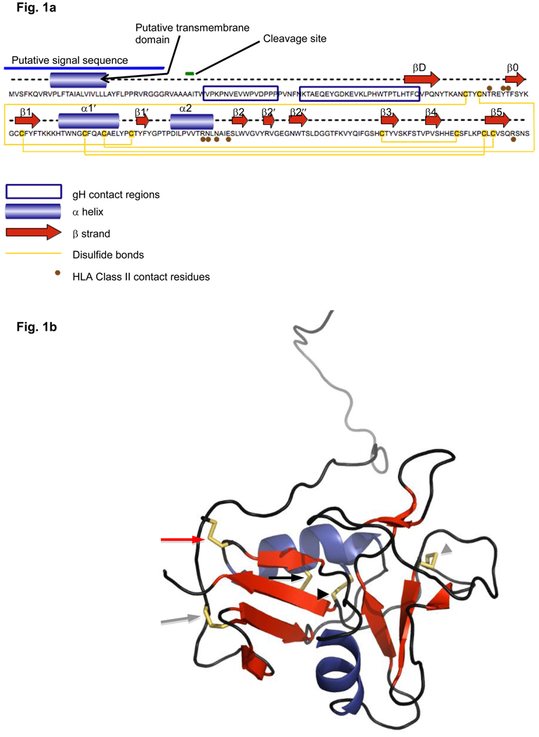
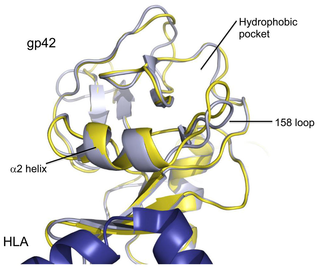
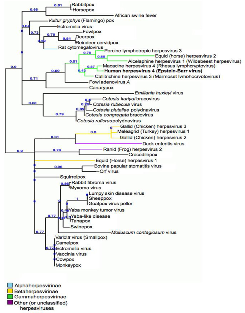
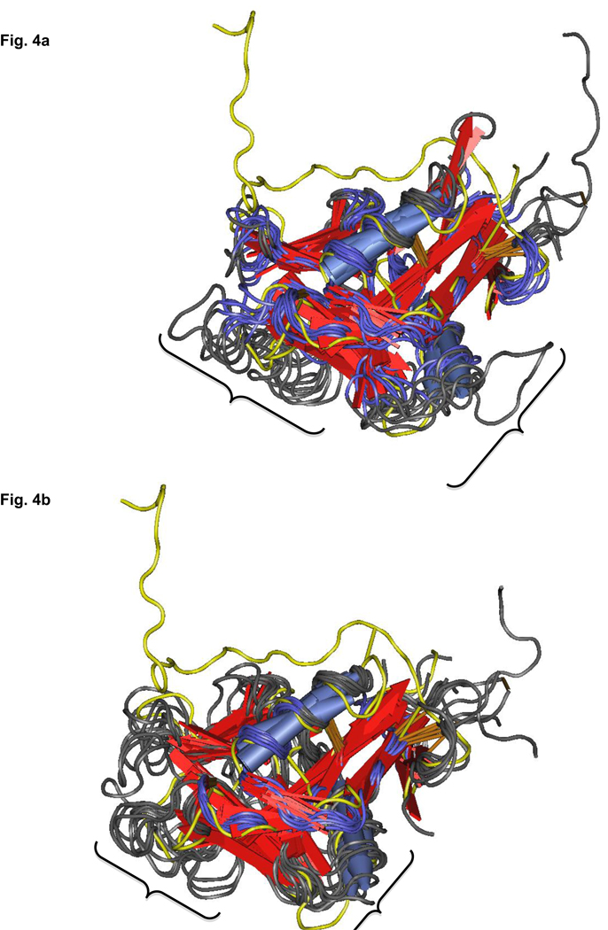
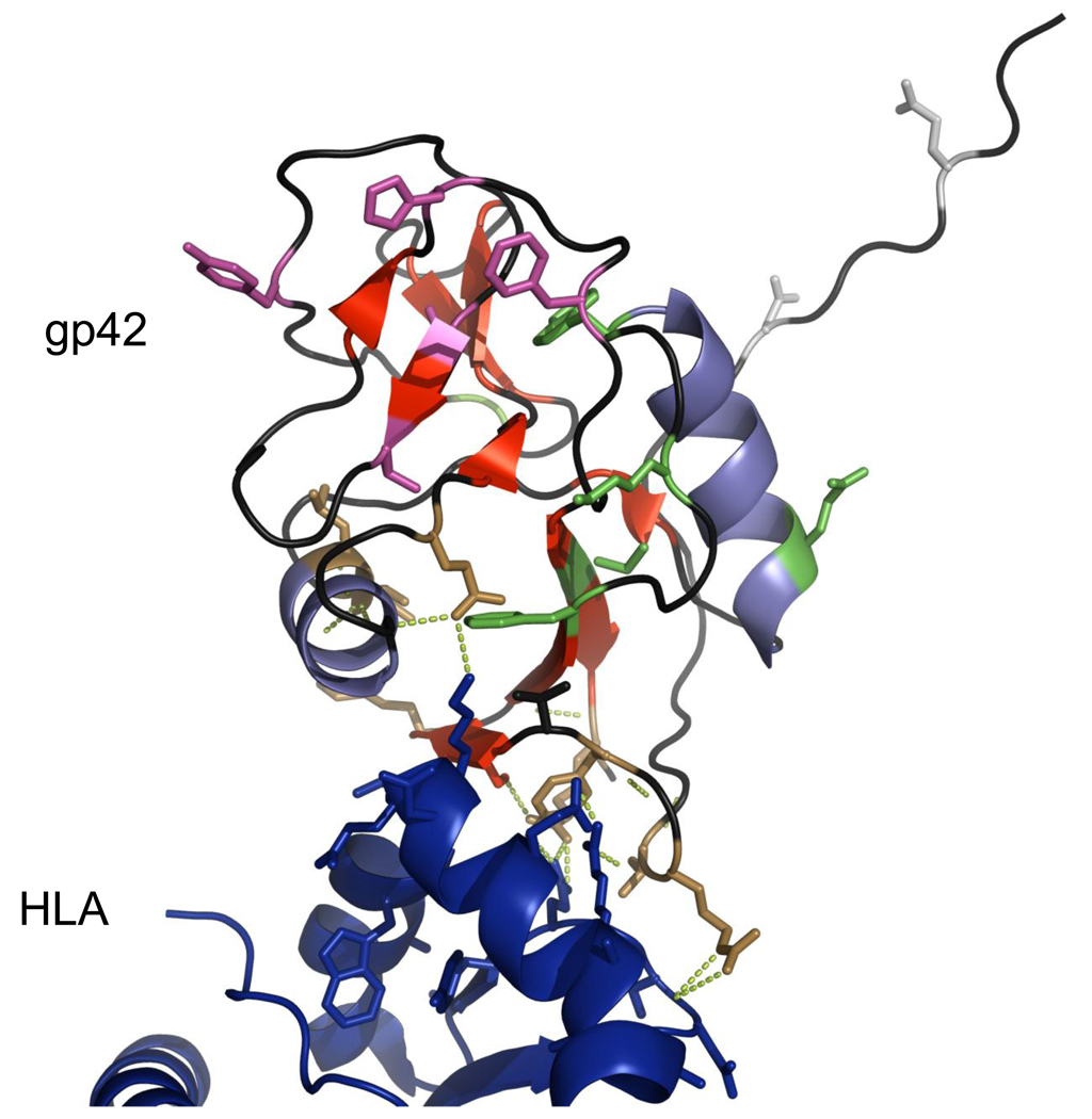
Epstein-Barr virus (EBV) glycoprotein 42 (gp42) is a membrane protein essential for fusion and entry of EBV into host B-lymphocytes. Gp42 is a member of the protein-fold family C-type lectin or lectin-like domains (CLECT or CTLD) and specifically is classified as a natural-killer receptor (NKR)-like CLECT. Literature review and phylogenetic comparison show that EBV gp42 shares a common structure with other NKR-like CLECTs and possibly with many viral CTLDs, but does not appear to exhibit some common binding characteristics of many CTLDs, such as features required for calcium binding. The flexible N-terminal region adjacent to the CTLD fold is important for binding to other EBV glycoproteins and for a cleavage site that is necessary for infection of host cells. From structural studies of gp42 unbound and bound to receptor and extensive mutational analysis, a general model of how gp42 triggers membrane fusion utilizing both the flexible N-terminal region and the CTLD domain has emerged.


Figures

Fig. 1
Fig. 1
Fig. 1a Linear graphical representation of the sequence and structural features of EBV gp42 Fig. 1b Corresponding x-ray crystallization-determined structure of gp42 in its unbound form beginning with amino acid 33 (Protein DataBank identification number 3FD4). Blue coils=alpha helices; red arrows=beta strands; yellow tubeworms=disulfide bridges; black arrowhead=disulfide bridge conserved in all canonical c-type lectin-like domains (CTLDs); gray arrowhead=disulfide bridge conserved in long-loop CTLDs; black arrow=disulfide bridge conserved in Ly49 natural killer (NK) CTLDs; gray arrow=disulfide bridge conserved in CD94 and NK2GD CTLD families; red arrow=disulfide bridge unique to EBV gp42. Figure created with Pymol
Fig. 2
Fig. 2
Overlay of the HLA class II-bound and unbound structures of EBV gp42. Bound structure is light blue-gray, unbound is yellow, HLA class II is deep blue. Figure created with Pymol
Fig. 3
Fig. 3
Rooted phylogenetic cladogram of viral CTLDs. Gamma herpesvirus CTLDs are grouped (green branch). Tree visualized with TreeDyn
Fig. 4
Fig. 4
Fig. 4a Nearest structural neighbors of EBV gp42 as determined by number of residues aligned with VAST. EBV gp42 is highlighted in yellow. Areas demarcated by brackets show less-aligned loops. Blue=alpha helices; red arrows=beta strands. Structures visualized with Cn3D Fig. 4b Re-alignment of nearest structural neighbors using the Loop-Hausdorff method with VAST. Loops are now more compactly aligned. Colors as in Fig. 4a. Structures visualized with Cn3D
Fig. 5
Fig. 5
Structure of EBVgp42 bound to HLA class II (PDB identification number 1KG0) with residues selected for mutation highlighted in color. Amino acids in the N-terminal domain are shown in silver-gray; HLA class II contacting residues are brown; hydrophobic pocket residues are pink-purple; residues in other structural features are green. Hydrogen bonds between selected gp42 and HLA contact residues are shown by chartreuse dotted lines. Structure visualized with Pymol

Similar articles

Cited by

References

    1. Rickinson A, Kieff E. In: Fields' Virology. Fields BN, Knipe DM, Howley PM, editors. Philadelphia: Lippincott Williams & Wilkins; 2007. pp. 2656–2700.
    1. Henle W, Henle G. Ann Clin Lab Sci. 1974;4:109–114. - PubMed
    1. Kutok JL, Wang F. Annu Rev Pathol. 2006;1:375–404. - PubMed
    1. Maeda E, Akahane M, Kiryu S, Kato N, Yoshikawa T, Hayashi N, Aoki S, Minami M, Uozaki H, Fukayama M, Ohtomo K. Jpn J Radiol. 2009;27:4–19. - PubMed
    1. Rezk SA, Weiss LM. Hum Pathol. 2007;38:1293–1304. - PubMed

Publication types