Antigenic strength controls the generation of antigen-specific IL-10-secreting T regulatory cells
- PMID: 20162554
- PMCID: PMC3466465
- DOI: 10.1002/eji.200940151
Antigenic strength controls the generation of antigen-specific IL-10-secreting T regulatory cells
Abstract
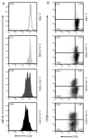
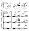
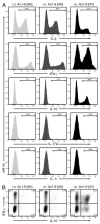
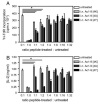
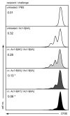
Administration of peptides i.n. induces peripheral tolerance in Tg4 myelin basic protein-specific TCR-Tg mice. This is characterized by the generation of anergic, IL-10-secreting CD4+ T cells with regulatory function (IL-10 Treg). Myelin basic protein Ac1-9 peptide analogs, displaying a hierarchy of affinities for H-2 A(u) (Ac1-9[4K]<<[4A]<[4Y]), were used to investigate the mechanisms of tolerance induction, focusing on IL-10 Treg generation. Repeated i.n. administration of the highest affinity peptide, Ac1-9[4Y], provided complete protection against EAE, while i.n. Ac1-9[4A] and Ac1-9[4K] treatment resulted in only partial protection. Ac1-9[4Y] was also the most potent stimulus for IL-10 Treg generation. Although i.n. treatment with Ac1-9[4A] gave rise to IL-10-secreting CD4+ T cells, the population as a whole was also capable of secreting IFN-gamma after an in vitro recall response to Ac1-9[4A] or [4Y]. In addition to IL-10 production, other facets of tolerance, namely, anergy and suppression (both in vitro and in vivo), were affinity dependent, with i.n. Ac1-9[4Y]-, [4A]- or [4K]-treated CD4+ T cells being the most, intermediate and least anergic/suppressive, respectively. These findings demonstrate that the generation of IL-10 Treg in vivo is driven by high signal strength.
Figures

Similar articles
-
Presentation of the self antigen myelin basic protein by dendritic cells leads to experimental autoimmune encephalomyelitis.J Immunol. 1999 Jul 1;163(1):32-9. J Immunol. 1999. PMID: 10384096
-
Inactivation of T cell receptor peptide-specific CD4 regulatory T cells induces chronic experimental autoimmune encephalomyelitis (EAE).J Exp Med. 1996 Nov 1;184(5):1609-17. doi: 10.1084/jem.184.5.1609. J Exp Med. 1996. PMID: 8920851 Free PMC article.
-
Peptide-induced T cell regulation of experimental autoimmune encephalomyelitis: a role for IL-10.Int Immunol. 1999 Oct;11(10):1625-34. doi: 10.1093/intimm/11.10.1625. Int Immunol. 1999. PMID: 10508180
-
Immune tolerance in multiple sclerosis.Immunol Rev. 2011 May;241(1):228-40. doi: 10.1111/j.1600-065X.2011.01016.x. Immunol Rev. 2011. PMID: 21488900 Free PMC article. Review.
-
Balancing autoaggressive and protective T cell responses.J Autoimmun. 2007 Mar-May;28(2-3):59-61. doi: 10.1016/j.jaut.2007.02.002. Epub 2007 Mar 23. J Autoimmun. 2007. PMID: 17363216 Free PMC article. Review.
Cited by
-
Controlling immune response and demyelination using highly potent bifunctional peptide inhibitors in the suppression of experimental autoimmune encephalomyelitis.Clin Exp Immunol. 2013 Apr;172(1):23-36. doi: 10.1111/cei.12029. Clin Exp Immunol. 2013. PMID: 23480182 Free PMC article.
-
Dietary supplementation with spray-dried animal plasma improves vaccine protection in aged mice.Front Nutr. 2023 Mar 24;10:1050961. doi: 10.3389/fnut.2023.1050961. eCollection 2023. Front Nutr. 2023. PMID: 37032769 Free PMC article.
-
Extra-thymically induced T regulatory cell subsets: the optimal target for antigen-specific immunotherapy.Immunology. 2015 Jun;145(2):171-81. doi: 10.1111/imm.12458. Immunology. 2015. PMID: 25716063 Free PMC article. Review.
-
Combination peptide immunotherapy suppresses antibody and helper T-cell responses to the RhD protein in HLA-transgenic mice.Haematologica. 2014 Mar;99(3):588-96. doi: 10.3324/haematol.2012.082081. Epub 2014 Jan 17. Haematologica. 2014. PMID: 24441145 Free PMC article.
-
Regulatory T Cell-Mediated Suppression of Inflammation Induced by DR3 Signaling Is Dependent on Galectin-9.J Immunol. 2017 Oct 15;199(8):2721-2728. doi: 10.4049/jimmunol.1700575. Epub 2017 Sep 6. J Immunol. 2017. PMID: 28877989 Free PMC article.
References
-
- Metzler B, Wraith DC. Inhibition of experimental autoimmune encephalomyelitis by inhalation but not oral administration of the encephalitogenic peptide: influence of MHC binding affinity. Int. Immunol. 1993;5:1159–1165. - PubMed
-
- Mason K, Denney DW, Jr., McConnell HM. Myelin basic peptide complexes with class II MHC molecules I-Au and I-Ak form and dissociate rapidly at neutral pH. J. Immunol. 1995;154:5216–5227. - PubMed
-
- Liu GY, Fairchild PJ, Smith RM, Prowle JR, Kioussis D, Wraith DC. Low avidity recognition of self-antigen by T cells permits escape from central tolerance. Immunity. 1995;3:407–415. - PubMed
-
- Burkhart C, Liu GY, Anderton SM, Metzler B, Wraith DC. Peptide-induced T cell regulation of experimental autoimmune encephalomyelitis: a role for IL-10. Int. Immunol. 1999;11:1625–1634. - PubMed
-
- Anderson PO, Sundstedt A, Yazici Z, Minaee S, Woolf R, Nicolson K, Whitley N, et al. IL-2 overcomes the unresponsiveness but fails to reverse the regulatory function of antigen-induced T regulatory cells. J. Immunol. 2005;174:310–319. - PubMed
Publication types
MeSH terms
Substances
Grants and funding
LinkOut - more resources
Full Text Sources
Research Materials
Miscellaneous

