. 2010 Jan;31(4):599-610.
doi: 10.1002/elps.200900441.

Identification of candidate biomarkers in ovarian cancer serum by depletion of highly abundant proteins and differential in-gel electrophoresis

Affiliations

Identification of candidate biomarkers in ovarian cancer serum by depletion of highly abundant proteins and differential in-gel electrophoresis

John D Andersen et al. Electrophoresis. 2010 Jan.

Abstract

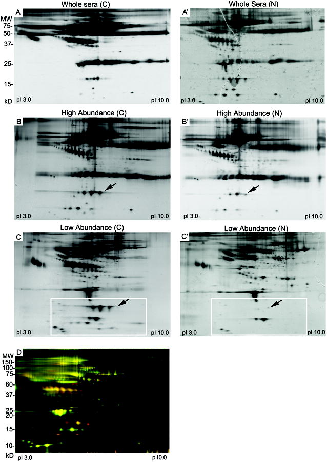
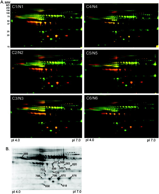
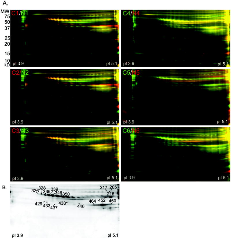
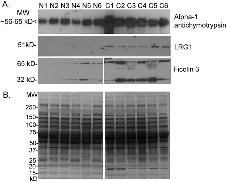
Ovarian cancer is the fifth leading cause of cancer death for women in the US, yet survival rates are over 90% when it is diagnosed at an early stage, highlighting the need for biomarkers for early detection. To enhance the discovery of tumor-specific proteins that could represent novel serum biomarkers for ovarian cancer, we depleted serum of highly abundant proteins which can mask the detection of proteins present in serum at low concentrations. Three commercial immunoaffinity columns were used in parallel to deplete the highly abundant proteins in serum from 60 patients with serous ovarian carcinoma and 60 non-cancer controls. Medium and low abundance serum proteins from each serum pool were then evaluated by the quantitative proteomic technique of differential in-gel electrophoresis. The number of protein spots that were elevated in ovarian cancer sera by at least twofold ranged from 36 to 248, depending upon the depletion and separation methods. From the 33 spots picked for MS analysis, nine different proteins were identified, including the novel candidate ovarian cancer biomarkers leucine-rich alpha2 glycoprotein-1 and ficolin 3. Western blotting validated the relative increases in serum protein levels for three of the proteins identified, demonstrating the utility of this approach for the identification of novel serum biomarkers for ovarian cancer.


Conflict of interest statement

Conflict of Interest Statement: The authors have declared no conflict of interest.

Figures

Figure 1
Figure 1. Depletion of highly abundant proteins from sera by MARS column
A) 2-DE of whole sera. Fifty μg of undepleted sera from pooled cancer (A) and non-cancer (A′) samples were separated by isoelectric point and then by molecular size. Proteins were visualized by silver stain. B) 2-DE of sera fractionated by the MARS column. Fifty μg of the high abundance proteins from cancer (B) and non-cancer (B′) serum pools were separated by 2-DE as described above. C) Fifty μg of the medium and low abundance proteins recovered from the MARS column for one cancer serum pool (C) and non-cancer serum pool (C′) were separated by 2-DE. Box indicates a region of the gel with protein spots of increased intensity in the ovarian cancer sera. Arrows show haptoglobin subunits. D) DIGE analysis of medium and low abundance proteins from sera depleted by the MARS column. Fifty μg of medium and low abundance proteins from ovarian cancer serum pool C1 was labeled with Cy5 (red) and 50 μg of medium and low abundance proteins from non-cancer serum pool N1 was labeled with Cy3 (green). Proteins present equally in both serum pools appear as yellow spots.
Figure 2
Figure 2. DIGE analysis of medium and low abundance serum proteins depleted by the IgY-12 spin column
A) DIGE gels for all 12 serum pools depleted of high abundance proteins by the IgY-12 spin column. Fifty μg of the medium and low abundance proteins recovered from the IgY-12 spin column were labeled with Cy5 (red; cancer serum pools C1-C6) or Cy3 (green; non-cancer serum pools N1-N6). Proteins present equally in both serum pools appear as yellow spots. Proteins were separated by isoelectric point and then by molecular size. B) A representative deep purple stained DIGE gel of sera depleted with the IgY-12 spin column. Spots picked for identification by MS are labeled with the master spot number assigned by the Decyder software. Identity of the proteins for each spot is listed in Table 2. The peptides identified for each protein are listed in Supplemental Table 1.
Figure 3
Figure 3. DIGE analysis of medium and low abundance serum proteins depleted by the IgY-12 HPLC column
A) DIGE gels for all 12 serum pools depleted of high abundance proteins by the IgY-12 HPLC column. Fifty μg of the medium and low abundance proteins recovered from the IgY-12 HPLC column were label for each serum pool. Cancer serum pools C1-C3 were labeled with Cy5 (red), and C4-C6 were labeled with Cy3 (green). Non-cancer serum pools N1-N3 were labeled with Cy3 (green), and N4-N6 were labeled with Cy5 (red). Proteins present equally in both serum pools appear as yellow spots. Proteins were separated by isoelectric point and then by molecular size. B) A representative deep purple stained DIGE gel of sera depleted with the IgY-12 HPLC column. Spots picked for identification by MS are labeled with the master spot number assigned by the Decyder software. The identity of the proteins for each spot is listed in Table 3. The peptides identified for each protein are listed in Supplemental Table 1.
Figure 4
Figure 4. Western blot validation of proteins overexpressed in cancer sera
A) Western blots of 50 μg of medium and low abundance proteins from each non-cancer (N1-N6) and cancer (C1-C6) serum pool depleted by the IgY-12 HPLC column were probed with the antibodies to AACT, LRG1, and ficolin 3. B) A duplicate gel was stained with CBB as a loading control.

References

    1. Jemal A, Siegel R, Ward E, Hao Y, Xu J, Murray T, Thun MJ. CA Cancer J Clin. 2008;58:71–96. - PubMed
    1. Jacobs IJ, Menon U. Mol Cell Proteomics. 2004;3:355–366. - PubMed
    1. Bast RC, Jr, Klug TL, St John E, Jenison E, Niloff JM, Lazarus H, Berkowitz RS, et al. N Engl J Med. 1983;309:883–887. - PubMed
    1. Bast RC, Jr, Urban N, Shridhar V, Smith D, Zhang Z, Skates S, Lu K, et al. Cancer Treat Res. 2002;107:61–97. - PubMed
    1. Zurawski VR, Jr, Orjaseter H, Andersen A, Jellum E. Int J Cancer. 1988;42:677–680. - PubMed

Publication types