

Dynamic EEG-informed fMRI modeling of the pain matrix using 20-ms root mean square segments
- PMID: 20162596
- PMCID: PMC6871058
- DOI: 10.1002/hbm.20967
Dynamic EEG-informed fMRI modeling of the pain matrix using 20-ms root mean square segments
Abstract
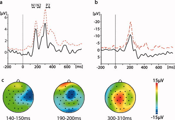
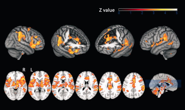
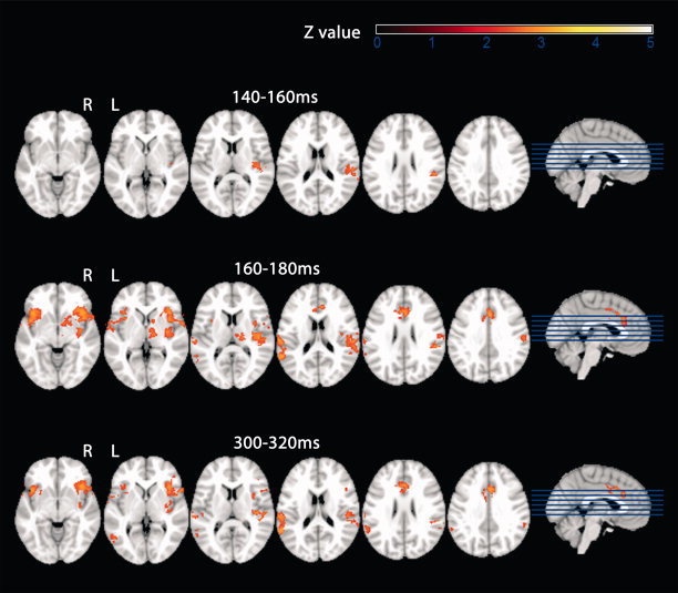
Previous studies on the spatio-temporal dynamics of cortical pain processing using electroencephalography (EEG), magnetoencephalography (MEG), or intracranial recordings point towards a high degree of parallelism, e.g. parallel instead of sequential activation of primary and secondary somatosensory areas or simultaneous activation of somatosensory areas and the mid-cingulate cortex. However, because of the inverse problem, EEG and MEG provide only limited spatial resolution and certainty about the generators of cortical pain-induced electromagnetic activity, especially when multiple sources are simultaneously active. On the other hand, intracranial recordings are invasive and do not provide whole-brain coverage. In this study, we thought to investigate the spatio-temporal dynamics of cortical pain processing in 10 healthy subjects using simultaneous EEG/functional magnetic resonance imaging (fMRI). Voltages of 20 ms segments of the EEG root mean square (a global, largely reference-free measure of event-related EEG activity) in a time window 0-400 ms poststimulus were used to model trial-to-trial fluctuations in the fMRI blood oxygen level dependent (BOLD) signal. EEG-derived regressors explained additional variance in the BOLD signal from 140 ms poststimulus onward. According to this analysis, the contralateral parietal operculum was the first cortical area to become activated upon painful laser stimulation. The activation pattern in BOLD analyses informed by subsequent EEG-time windows suggests largely parallel signal processing in the bilateral operculo-insular and mid-cingulate cortices. In that regard, our data are in line with previous reports. However, the approach presented here is noninvasive and bypasses the inverse problem using only temporal information from the EEG.
© 2010 Wiley-Liss, Inc.
Figures
Similar articles
-
Fluctuations in electrodermal activity reveal variations in single trial brain responses to painful laser stimuli--a fMRI/EEG study.Neuroimage. 2009 Feb 1;44(3):1081-92. doi: 10.1016/j.neuroimage.2008.09.004. Epub 2008 Sep 20. Neuroimage. 2009. PMID: 18848631
-
Laser-evoked potential P2 single-trial amplitudes covary with the fMRI BOLD response in the medial pain system and interconnected subcortical structures.Neuroimage. 2009 Apr 15;45(3):917-26. doi: 10.1016/j.neuroimage.2008.12.051. Epub 2009 Jan 7. Neuroimage. 2009. PMID: 19166948
-
Integration of EEG source imaging and fMRI during continuous viewing of natural movies.Magn Reson Imaging. 2010 Oct;28(8):1135-42. doi: 10.1016/j.mri.2010.03.042. Epub 2010 Jun 25. Magn Reson Imaging. 2010. PMID: 20579829
-
Functional imaging of brain responses to pain. A review and meta-analysis (2000).Neurophysiol Clin. 2000 Oct;30(5):263-88. doi: 10.1016/s0987-7053(00)00227-6. Neurophysiol Clin. 2000. PMID: 11126640 Review.
-
Brain generators of laser-evoked potentials: from dipoles to functional significance.Neurophysiol Clin. 2003 Dec;33(6):279-92. doi: 10.1016/j.neucli.2003.10.008. Neurophysiol Clin. 2003. PMID: 14678842 Review.
Cited by
-
Ketamine effects on default mode network activity and vigilance: A randomized, placebo-controlled crossover simultaneous fMRI/EEG study.Hum Brain Mapp. 2020 Jan;41(1):107-119. doi: 10.1002/hbm.24791. Epub 2019 Sep 18. Hum Brain Mapp. 2020. PMID: 31532029 Free PMC article. Clinical Trial.
-
High-Density Electroencephalography-Informed Multiband Functional Magnetic Resonance Imaging Reveals Rhythm-Specific Activations Within the Trigeminal Nociceptive Network.Front Neurosci. 2022 May 16;16:802239. doi: 10.3389/fnins.2022.802239. eCollection 2022. Front Neurosci. 2022. PMID: 35651631 Free PMC article.
-
Neurological diseases and pain.Brain. 2012 Feb;135(Pt 2):320-44. doi: 10.1093/brain/awr271. Epub 2011 Nov 8. Brain. 2012. PMID: 22067541 Free PMC article. Review.
References
-
- Allen P, Polizzi G, Krakow K, Fish DR, Lemieux L ( 1998): Identification of EEG Events in the MR scanner: The problem of pulse artifact and a method for its subtraction. NeuroImage 8: 229–239. - PubMed
-
- Allen P, Josephs O, Turner R ( 2000): A method for removing imaging artifact from continuous EEG recorded during functional MRI. NeuroImage 1: 230–239. - PubMed
-
- Apkarian AV, Bushnell MC, Treede RD, Zubieta JK ( 2005): Human brain mechanisms of pain perception and regulation in health and disease. Eur J Pain 9: 463–484. - PubMed
-
- Baudena P, Halgren E, Heit G, Clarke JM ( 1995): Intracerebral potentials to rare target and distractor auditory and visual stimuli. III. Frontal cortex. Electroencephalogr Clin Neurophysiol 94: 251–264. - PubMed
-
- Behrens T, Woolrich MW, Smith S ( 2003): Multi‐Testing Using a Fully Subject Null Hypothesis Bayesian Framework: Theory. New York: Human Brain Mapping Meeting.
Publication types
MeSH terms
LinkOut - more resources
Full Text Sources
Medical
