

Abnormal water diffusivity in corticostriatal projections in children with Tourette syndrome
- PMID: 20162597
- PMCID: PMC6871238
- DOI: 10.1002/hbm.20970
Abnormal water diffusivity in corticostriatal projections in children with Tourette syndrome
Abstract
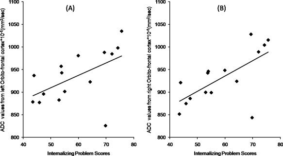
The fronto-striato-thalamic circuit has been implicated in the pathomechanism of Tourette Syndrome (TS). To study white and gray matter comprehensively, we used a novel technique called Tract-Based Spatial Statistics (TBSS) combined with voxel-based analysis (VBA) of diffusion tensor MR images in children with TS as compared to typically developing controls. These automated and unbiased methods allow analysis of cerebral white matter and gray matter regions. We compared 15 right-handed children with TS (mean age: 11.6 ± 2.5 years, 12 males) to 14 age-matched right-handed healthy controls (NC; mean age: 12.29 ± 3.2 years, 6 males). Tic severity and neurobehavioral scores were correlated with FA and ADC values in regions found abnormal by these methods. For white matter, TBSS analysis showed regions of increased ADC in the corticostriatal projection pathways including left external capsule and left and right subcallosal fasciculus pathway in TS group compared to NC group. Within the TS group, ADC for the left external capsule was negatively associated with tic severity (r= -0.586, P = 0.02). For gray matter, VBA revealed increased ADC for bilateral orbitofrontal cortex, left putamen, and left insular cortex. ADC for the right and left orbitofrontal cortex was highly correlated with internalizing problems (r = 0.665; P = 0.009, r = 0.545; P = 0.04, respectively). Altogether, this analysis revealed focal diffusion abnormalities in the corticostriatal pathway and in gray matter structures involved in the fronto-striatal circuit in TS. These diffusion abnormalities could serve as a neuroimaging marker related to tic severity and neurobehavioral abnormalities in TS subjects.
© 2010 Wiley-Liss, Inc.
Figures


Similar articles
-
Combining tract- and atlas-based analysis reveals microstructural abnormalities in early Tourette syndrome children.Hum Brain Mapp. 2016 May;37(5):1903-19. doi: 10.1002/hbm.23146. Epub 2016 Mar 1. Hum Brain Mapp. 2016. PMID: 26929221 Free PMC article.
-
Microstructural abnormalities of striatum and thalamus in children with Tourette syndrome.Mov Disord. 2008 Dec 15;23(16):2349-56. doi: 10.1002/mds.22264. Mov Disord. 2008. PMID: 18759338
-
Characteristics of diffusion tensor imaging of central nervous system in children with tourette's disease.Medicine (Baltimore). 2020 May 29;99(22):e20492. doi: 10.1097/MD.0000000000020492. Medicine (Baltimore). 2020. PMID: 32481462 Free PMC article.
-
Cingulate role in Tourette syndrome.Handb Clin Neurol. 2019;166:165-221. doi: 10.1016/B978-0-444-64196-0.00011-X. Handb Clin Neurol. 2019. PMID: 31731911 Review.
-
The role of diffusion tensor imaging and fractional anisotropy in the evaluation of patients with idiopathic normal pressure hydrocephalus: a literature review.Neurosurg Focus. 2016 Sep;41(3):E12. doi: 10.3171/2016.6.FOCUS16192. Neurosurg Focus. 2016. PMID: 27581308 Review.
Cited by
-
Altered frontal-mediated inhibition and white matter connectivity in pediatric chronic tic disorders.Exp Brain Res. 2021 Mar;239(3):955-965. doi: 10.1007/s00221-020-06017-0. Epub 2021 Jan 18. Exp Brain Res. 2021. PMID: 33462641 Free PMC article.
-
Compensatory neural reorganization in Tourette syndrome.Curr Biol. 2011 Apr 12;21(7):580-5. doi: 10.1016/j.cub.2011.02.047. Curr Biol. 2011. PMID: 21439830 Free PMC article.
-
Alterations in the microstructure of white matter in children and adolescents with Tourette syndrome measured using tract-based spatial statistics and probabilistic tractography.Cortex. 2018 Jul;104:75-89. doi: 10.1016/j.cortex.2018.04.004. Epub 2018 Apr 12. Cortex. 2018. PMID: 29758375 Free PMC article.
-
Diffuse alterations in grey and white matter associated with cognitive impairment in Shwachman-Diamond syndrome: evidence from a multimodal approach.Neuroimage Clin. 2015 Feb 27;7:721-31. doi: 10.1016/j.nicl.2015.02.014. eCollection 2015. Neuroimage Clin. 2015. PMID: 25844324 Free PMC article.
-
ChangPu YuJin Tang improves Tourette disorder symptoms by modulating amino acid neurotransmitters in IDPN model rats.Metab Brain Dis. 2024 Dec;39(8):1543-1558. doi: 10.1007/s11011-024-01411-x. Epub 2024 Sep 23. Metab Brain Dis. 2024. PMID: 39312065
References
-
- Ashburner J, Andersson JL, Friston KJ ( 1999): High‐dimensional image registration using symmetric priors. Neuroimage 9: 619–628. - PubMed
-
- Ashburner J, Friston KJ ( 2000): Voxel‐based morphometry—The methods. Neuroimage 11: 805–821. - PubMed
-
- Basser PJ, Mattiello J, LeBihan D ( 1994): Estimation of the effective self‐diffusion tensor from the NMR spin echo. J Magn Reson B 103: 247–254. - PubMed
-
- Behen M, Chugani HT, Juhasz C, Helder E, Ho A, Maqbool M, Rothermel RD, Perry J, Muzik O ( 2007): Abnormal brain tryptophan metabolism and clinical correlates in Tourette syndrome. Mov Disord 22: 2256–2262. - PubMed
