

Assessment of white matter tract damage in mild cognitive impairment and Alzheimer's disease
- PMID: 20162601
- PMCID: PMC6870900
- DOI: 10.1002/hbm.20978
Assessment of white matter tract damage in mild cognitive impairment and Alzheimer's disease
Abstract
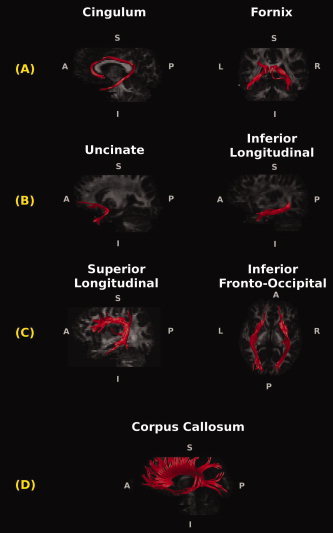
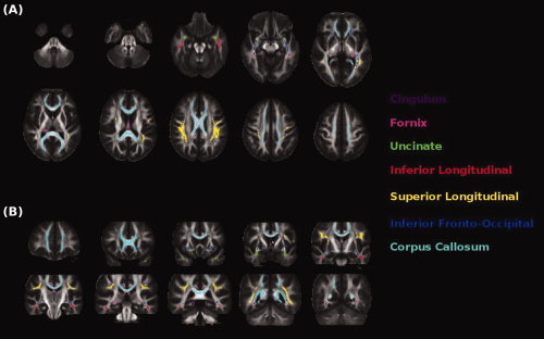
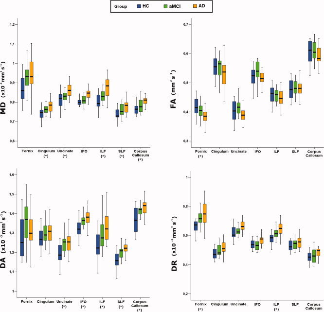
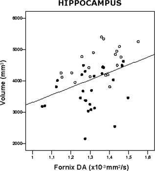
Diffusion tensor MRI-based tractography was used to investigate white matter (WM) changes in the major limbic (i.e., fornix and cingulum) and cortico-cortical association pathways [i.e., the uncinate fasciculus, the inferior fronto-occipital fasciculus, the inferior longitudinal fasciculus (ILF), the superior longitudinal fasciculus, and the corpus callosum] in 25 Alzheimer's disease (AD) patients, 19 amnestic mild cognitive impairment (aMCI) patients, and 15 healthy controls (HC). Mean diffusivity (MD), fractional anisotropy (FA), as well as axial (DA) and radial (DR) diffusivities were measured for each tract, using an atlas-based tractography approach. The association of WM tract integrity with hippocampal volume was also assessed. MD values were significantly different among groups in all WM tracts (P values ranging from 0.002 to 0.03), except in the fornix (P = 0.06) and the inferior fronto-occipital fasciculus (P = 0.09). Conversely, FA was significantly different among groups in the fornix only (P = 0.02). DA values were significantly different among groups in all WM tracts (P values ranging from 0.001 to 0.01), except in the fornix (P = 0.13) and the cingulum (P = 0.29). Significantly different DR values among groups were found in the fornix (P = 0.02) and the ILF (P = 0.01). In the fornix and cingulum, DR was significantly more increased than DA in both patient groups compared to HC. No difference in DA versus DR was found in cortico-cortical WM tracts. DA values in the fornix were significantly correlated with the hippocampal volume. This study demonstrates a different pattern of WM involvement in the limbic and cortico-cortical association pathways in aMCI and AD patients.
Copyright © 2010 Wiley-Liss, Inc.
Figures
Similar articles
-
Distinctive disruption patterns of white matter tracts in Alzheimer's disease with full diffusion tensor characterization.Neurobiol Aging. 2012 Sep;33(9):2029-45. doi: 10.1016/j.neurobiolaging.2011.06.027. Epub 2011 Aug 27. Neurobiol Aging. 2012. PMID: 21872362 Free PMC article.
-
Microstructural white matter changes, not hippocampal atrophy, detect early amnestic mild cognitive impairment.PLoS One. 2013;8(3):e58887. doi: 10.1371/journal.pone.0058887. Epub 2013 Mar 14. PLoS One. 2013. PMID: 23516569 Free PMC article.
-
Comparison of diffusion tensor image study in association fiber tracts among normal, amnestic mild cognitive impairment, and Alzheimer's patients.Neurol India. 2011 Mar-Apr;59(2):168-73. doi: 10.4103/0028-3886.79129. Neurol India. 2011. PMID: 21483111
-
White matter integrity and vulnerability to Alzheimer's disease: preliminary findings and future directions.Biochim Biophys Acta. 2012 Mar;1822(3):416-22. doi: 10.1016/j.bbadis.2011.07.009. Epub 2011 Jul 23. Biochim Biophys Acta. 2012. PMID: 21803153 Free PMC article. Review.
-
Diffusion tensor imaging of white matter degeneration in early stage of Alzheimer's disease: a review.Int J Neurosci. 2020 Mar;130(3):243-250. doi: 10.1080/00207454.2019.1667798. Epub 2019 Sep 24. Int J Neurosci. 2020. PMID: 31549530
Cited by
-
Different patterns of white matter degeneration using multiple diffusion indices and volumetric data in mild cognitive impairment and Alzheimer patients.PLoS One. 2012;7(12):e52859. doi: 10.1371/journal.pone.0052859. Epub 2012 Dec 31. PLoS One. 2012. PMID: 23300797 Free PMC article.
-
Longitudinal, region-specific course of diffusion tensor imaging measures in mild cognitive impairment and Alzheimer's disease.Alzheimers Dement. 2013 Sep;9(5):519-28. doi: 10.1016/j.jalz.2012.05.2186. Epub 2012 Dec 12. Alzheimers Dement. 2013. PMID: 23245561 Free PMC article.
-
Assessing corpus callosum changes in Alzheimer's disease: comparison between tract-based spatial statistics and atlas-based tractography.PLoS One. 2012;7(4):e35856. doi: 10.1371/journal.pone.0035856. Epub 2012 Apr 24. PLoS One. 2012. PMID: 22545143 Free PMC article.
-
Staging neurodegenerative disorders: structural, regional, biomarker, and functional progressions.Neurotox Res. 2011 Feb;19(2):211-34. doi: 10.1007/s12640-010-9190-2. Neurotox Res. 2011. PMID: 20393891 Review.
-
An advanced white matter tract analysis in frontotemporal dementia and early-onset Alzheimer's disease.Brain Imaging Behav. 2016 Dec;10(4):1038-1053. doi: 10.1007/s11682-015-9458-5. Brain Imaging Behav. 2016. PMID: 26515192 Free PMC article.
References
-
- Amodio P, Wenin H, Del Piccolo F, Mapelli D, Montagnese S, Pellegrini A, Musto C, Gatta A, Umilta C ( 2002): Variability of trail making test, symbol digit test and line trait test in normal people. A normative study taking into account age‐dependent decline and sociobiological variables. Aging Clin Exp Res 14: 117–131. - PubMed
-
- Arnold SE, Hyman BT, Flory J, Damasio AR, Van Hoesen GW ( 1991): The topographical and neuroanatomical distribution of neurofibrillary tangles and neuritic plaques in the cerebral cortex of patients with Alzheimer's disease. Cereb Cortex 1: 103–116. - PubMed
-
- Barigazzi ( 1987): Esplorazione testistica della memoria di prosa. Ric Psicol 1: 50–80.
-
- Bartzokis G ( 2004): Age‐related myelin breakdown: A developmental model of cognitive decline and Alzheimer's disease. Neurobiol Aging 25: 5–18; author reply 49–62. - PubMed
-
- Basser PJ, Pierpaoli C ( 1996): Microstructural and physiological features of tissues elucidated by quantitative‐diffusion‐tensor MRI. J Magn Reson B 111: 209–219. - PubMed
Publication types
MeSH terms
LinkOut - more resources
Full Text Sources
Medical
