. 2010 Apr;19(4):836-46.
doi: 10.1002/pro.363.

Discovery and characterization of a mammalian amyloid disaggregation activity

Affiliations

Discovery and characterization of a mammalian amyloid disaggregation activity

Amber N Murray et al. Protein Sci. 2010 Apr.

Abstract

The formation of amyloid, a cross-beta-sheet fibrillar aggregate, is associated with a variety of aging-associated degenerative diseases. Herein, we report the existence of a mammalian amyloid disaggregase activity that is present in all tissues and cell types tested. Homogenates from mammalian tissues and cell lines are able to disaggregate amyloid fibrils composed of amyloid beta (A beta)(1-40) or the 8 kDa plasma gelsolin fragment. The mammalian disaggregase activity is sensitive to proteinase K digestion and can be uncoupled from proteolysis activity using a protease inhibitor cocktail. Amyloid disaggregation and proteolysis activities are remarkably resistant to changes in temperature and pH. Identification and manipulation of the proteins responsible for the amyloid disaggregation/degradation activities offers the possibility of ameliorating aggregation-associated diseases.


Figures

Figure 1
Figure 1
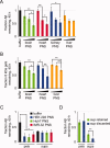
PNS from mouse tissues disaggregates amyloid fibrils. (Error bars indicate standard deviations in three independent disaggregation experiments; * denotes P < 0.05 and ** denotes P < 0.01). A: Mouse brain, heart, kidney, and liver PNS (31 μg/mL total protein) disaggregate Aβ1–40 fibrils (86 μg/mL), as compared with BSA (31 μg/mL) or buffer alone controls. B: Mouse brain, heart, kidney, and liver PNS (45 μg/mL total protein) disaggregate 8 kDa gelsolin fibrils (31 μg/mL), as compared with BSA (45 μg/mL) or buffer alone controls. C: The extent of Aβ1–40 fibril (65 μg/mL) disaggregation (measured at 40 h) depends on the total protein concentration of mouse tissue PNS (3–30 μg/mL total protein), with higher concentrations leading to more disaggregation. All samples have a statistically significantly lower fluorescence than the buffer alone control except for BSA and 3 μg/mL liver PNS, with P < 0.01. D: The extent of 8 kDa gelsolin fibril (31 μg/mL) disaggregation is similarly dependent on total PNS protein concentration (3–30 μg/mL total protein). All samples have a statistically significantly lower fluorescence than the buffer alone control except for BSA, with P < 0.01. E: AFM confirms a decrease in the amount of Aβ1–40 fibrils (65 μg/mL) following disaggregation by mouse heart PNS (30 μg/mL total protein) (bottom panels), when compared with a buffer alone control (top panels). F: AFM demonstrates a decrease in 8 kDa gelsolin fibrils (31 μg/mL) following disaggregation by mouse kidney PNS (30 μg/mL total protein) (bottom vs. top panels). In each case, a representative image of the fewest fibrils observed in the 10 or more fields examined is shown in the left panels, and a representative image of the most fibrils observed is shown in the right panels.
Figure 2
Figure 2
PNS-mediated amyloid disaggregase activity is sensitive to proteolytic digestion. Proteinase K (PK) (2 μg/mL) pretreatment of mouse kidney PNS (300 μg/mL total protein) eliminates its ability (at 10 μg/mL total protein) to disaggregate Aβ1–40 fibrils (65 μg/mL, blue), when compared with untreated control (green).
Figure 3
Figure 3
PNS from human cell lines disaggregates amyloid fibrils. (Error bars indicate standard deviations in three independent disaggregation experiments; * denotes p < 0.05 and ** denotes p < 0.01). A: HEK 293, Huh7, and IMR-32 PNS (30 μg/mL total protein) disaggregates Aβ1–40 fibrils (65 μg/mL), when compared with BSA (30 μg/mL) or buffer alone controls. B: HEK 293, Huh7, and IMR-32 PNS (30 μg/mL total protein) disaggregates 8 kDa gelsolin fibrils (31 μg/mL), as compared with BSA (30 μg/mL) or buffer alone controls. C: The extent of Aβ1–40 fibril (65 μg/mL) disaggregation (measured at 40 h) depends on the total protein concentration of human cell PNS (3–30 μg/mL total protein), with higher concentrations leading to more disaggregation. All samples have a statistically significantly lower fluorescence than the buffer alone control except for BSA, with P < 0.01. D: The extent of 8 kDa gelsolin disaggregation (31 μg/mL) also depends on the total protein concentration of human cell PNS (3–30 μg/mL total protein). All samples have a statistically significantly lower fluorescence than the buffer alone control except for BSA and 3 μg/mL Huh7 PNS, with P < 0.05. E: AFM confirms a decrease in the amount of Aβ1–40 fibrils (65 μg/mL) following disaggregation by Huh7 PNS (30 μg/mL total protein) (bottom panels), when compared with a buffer alone control (top panels). F: AFM demonstrates a decrease in 8 kDa gelsolin fibrils (31 μg/mL) following disaggregation by HEK 293 PNS (30 μg/mL total protein) (bottom vs. top panels). In each case, a representative image of the fewest fibrils observed in the 10 or more fields examined is shown in the left panels, and a representative image of the most fibrils observed is shown in the right panels.
Figure 4
Figure 4
Mammalian PNS degrades Aβ1–40 and 8 kDa gelsolin from fibrils, depleting high-molecular weight species. (Error bars indicate standard deviations in three independent proteolysis experiments; * denotes p < 0.05 and ** denotes p < 0.01.) A: Quantification of Aβ1–40 reversed phase liquid chromatography peak areas indicates that mouse tissue PNS degrades Aβ1–40 (65 μg/mL), and the extent of proteolysis is proportional to the concentration of total PNS protein (3–30 μg/mL total protein). All 10 and 30 μg/mL PNS samples have statistically significantly less Aβ1–40 remaining, with P < 0.05. B: Quantification of 8 kDa gelsolin reversed phase liquid chromatography peak areas shows that mouse tissue PNS (3–30 μg/mL total protein) also degrades 8 kDa gelsolin (31 μg/mL) in a dose-dependent fashion. All 10 and 30 μg/mL PNS samples have statistically significantly less gelsolin remaining, with P < 0.05. C: Human cell PNS (5 μg/mL total protein) depletes the pelletable (200,000 g for 1 h) population (pels) of Aβ1–40 but does not affect the supernatant Aβ1–40 population (sups). D: Removal of soluble Aβ1–40 following a 200,000 g spin at t = 0 (sup discarded, blue) does not repopulate the supernatant pool from a 200,000 g spin after a 40-h incubation period (cf. short blue bar, sups, to short green bar, sups).
Figure 5
Figure 5
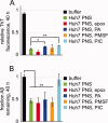
Mammalian amyloid disaggregation and proteolysis activities are uncoupled by a protease inhibitor cocktail. (Error bars indicate standard deviations in three independent disaggregation or proteolysis experiments; * denotes p < 0.05 and ** denotes p < 0.01. A: Phosphoramidon (PA, 20 μM) and Roche Complete EDTA-Free Protease Inhibitor Cocktail (PIC, at 1 tablet per 50 mL of solution) slightly reduce disaggregation of Aβ1–40 fibrils (65 μg/mL) by Huh7 PNS (30 μg/mL total protein), increasing the average relative ThT fluorescence at 40 h from 0.14 (Huh7 PNS, green) to 0.19 (Huh7 PNS, PA, blue) and 0.16 (Huh7 PNS, PIC, cyan). Epoxomicin (epox, 10 μM) and PMSF (1 mM) do not significantly affect disaggregation of Aβ1–40 fibrils by Huh7 PNS. All samples have a statistically significantly lower fluorescence than the buffer alone control, with P < 0.01. B: Only Roche Complete EDTA-Free Protease Inhibitor Cocktail (PIC, cyan) prevents Huh7 PNS-mediated Aβ1–40 proteolysis under the same conditions.
Figure 6
Figure 6
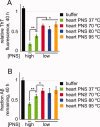
Mammalian amyloid disaggregase and proteolysis activities are resistant to high-temperature pretreatment. (Error bars indicate standard deviations in three independent disaggregation or proteolysis experiments; * denotes p < 0.05 and ** denotes p < 0.01.) A: Pretreatment (2 h) of mouse heart PNS (10 μg/mL, low) at temperatures up to 95 °C decreases disaggregation of Aβ1–40 fibrils (65 μg/mL) but does not completely inhibit the disaggregase activity. There is a statistically significantly lower ThT signal from fibrils to which a higher concentration of PNS was added (10 μg/mL, high), corroborating that the ratio of 65 μg/mL Aβ1–40 fibrils: 10 μg/mL mouse heart PNS is within the linear range of the disaggregation assay. All samples have a statistically significantly lower fluorescence than the buffer alone control, with P < 0.01. B: Pretreatment (2 h) of mouse heart PNS (10 μg/mL) at temperatures up to 95 °C does not prevent proteolysis of Aβ1–40 (65 μg/mL). All samples have a statistically significantly lower Aβ1–40 peak area than the buffer alone control, with P < 0.05.
Figure 7
Figure 7
Mammalian amyloid disaggregase and proteolysis activities are sensitive to low and high pH pretreatment. Dialysis of HEK 293 PNS (1 mg/mL total protein) for 16 h at 4 °C into buffers at low pH (and to a lesser extent high pH) decreases its ability (at a final total PNS concentration of 10 μg/mL) to disaggregate Aβ1–40 fibrils (65 μg/mL) and to degrade Aβ1–40.

References

    1. Balch WE, Morimoto RI, Dillin A, Kelly JW. Adapting proteostasis for disease intervention. Science. 2008;319:916–919. - PubMed
    1. Powers ET, Morimoto RI, Dillin A, Kelly JW, Balch WE. Biological and chemical approaches to diseases of proteostasis deficiency. Annu Rev Biochem. 2009;78:959–991. - PubMed
    1. Hammarstrom P, Schneider F, Kelly JW. Trans-suppression of misfolding in an amyloid disease. Science. 2001;293:2459–2462. - PubMed
    1. Chen C-D, Huff ME, Matteson J, Page L, Phillips R, Kelly JW, Balch WE. Furin initiates gelsolin familial amyloidosis in the Golgi through a defect in Ca2+ stabilization. EMBO J. 2001;20:6277–6287. - PMC - PubMed
    1. Selkoe DJ. Folding proteins in fatal ways. Nature. 2003;426:900–904. - PubMed

Publication types