. 2010 Apr 6;11(5):1029-35.
doi: 10.1002/cphc.200900914.

Tailored porous silicon microparticles: fabrication and properties

Affiliations

Tailored porous silicon microparticles: fabrication and properties

Ciro Chiappini et al. Chemphyschem. .

Abstract

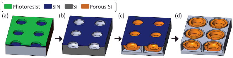
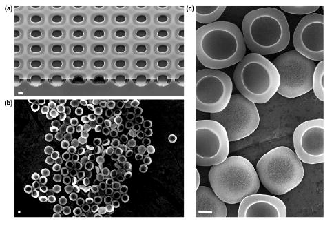
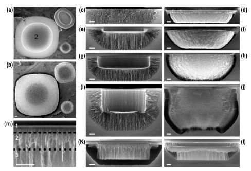
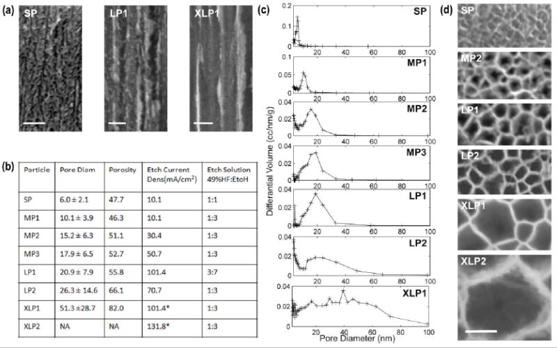
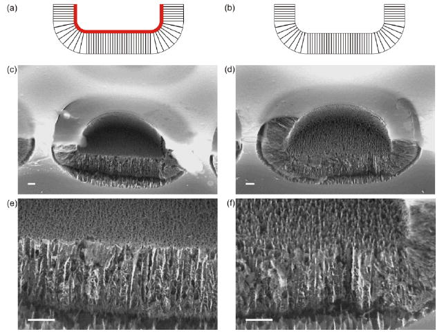
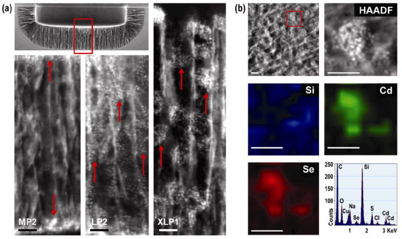
The use of mesoporous silicon particles for drug delivery has been widely explored thanks to their biodegradability and biocompatibility. The ability to tailor the physicochemical properties of porous silicon at the micro- and nanoscale confers versatility to this material. A method for the fabrication of highly reproducible, monodisperse, mesoporous silicon particles with controlled physical characteristics through electrochemical etching of patterned silicon trenches is presented. The particle size is tailored in the micrometer range and pore size in the nanometer range, the shape from tubular to discoidal to hemispherical, and the porosity from 46 to over 80%. In addition, the properties of the porous matrix are correlated with the loading of model nanoparticles (quantum dots) and their three-dimensional arrangement within the matrix is observed by transmission electron microscopy tomography. The methods developed in this study provide effective means to fabricate mesoporous silicon particles according to the principles of rational design for therapeutic vectors and to characterize the distribution of nanoparticles within the porous matrix.


Figures

Figure 1
Figure 1
Schematic depiction of the fabrication process. (a) Pattern transfer to the photoresist layer on top of the sacrificial SiN layer. (b) Trench formation in the Si substrate through combination of dry and wet etch. (c) Formation of the PSPs and release layer following anodic etch. (d) PSPs ready to be released by ultrasonication in isopropanol following stripping of the SiN mask
Figure 2
Figure 2
Scanning electron micrographs of large clusters of PSPs. The PSPs are characterized by size and shape uniformity. (a) HBr6 etched tubular shaped MP3 PSPs still attached to silicon substrate before the removal of the silicon nitride sacrificial layer and the PSPs subsequent release. (b) overview of a large cluster of SF6 etched Bowl shaped MP3 PSPs following release by sonication in IPA. (c) Close-up of a small cluster of CF4 etched discoidal shaped MP3 PSPs following release by sonication in IPA. All scale bars are 1 μm
Figure 3
Figure 3
SEM micrographs of PSPs. (a) Digital composition of three distinct SEM micrographs showing the nucleation side of a 3.2μm, 1.6 μm and 0.97 μm PSP: (1) the external corona and (2) the nucleation site. (b) Digital composition of three distinct SEM micrographs showing the release side of a 3.2μm, 1.6 μm and 0.97 μm PSP. Section along the diameter and the lateral view of: a flat disk PSP obtained by wet etch of the masking layer (c, d respectively); a discoidal PSP obtained by trench formation by CF4 RIE (e, f respectively); a hemispherical PSP obtained by trench formation by SF6 RIE (g, h respectively); a tubular PSP obtained by trench formation by combination of HBr and SF6 RIE (i, j respectively); a XLP1 PSP (k, l respectively). (m) Close-up view of the multilayer structure of an XLP1 particle, from top to bottom: (1) SP layer, (2) transitional layer, (3) XLP layer. (c-m) Nucleation side is at the top of the figure and release side at the bottom. All scale bars are 200nm.
Figure 4
Figure 4
Analysis of the porous structure of the PSPs. (a) SEM micrographs of the PSP porous structure cross section along the pore axis, perpendicular to the Si (100) surface for the most representative pore structures investigated. (b) Summary of the BJH analysis and the electrochemical etch parameters employed in the porosification of the PSPs. The etch current density reported for XLPs refers to the XLP layer. (c) BJH model estimate of the differential volume of pores in the 3nm to 100nm range for all the different types of PSPs investigated. BJH analysis of XLP2 PSPs is not shown due to the inadequacy of the model. (d) SEM micrographs of the central bottom region of the PSP for the most representative pore sizes investigated (all scale bars in the figure are100nm).
Figure 5
Figure 5
A short CF4 plasma etch following the formation of the PSP results in the removal of the nucleation layer and formation of pass-through pores. (a) Schematic depiction of a PSP as anodically etched; the small pore nucleation layer is indicated in red. (b) Schematic depticion of a PSP following 15s CF4 plasma etch. (c-f) 45° tilt SEM cross section of a PSP with (a,c) and without (b,d) nucleation layer. No damage to the PSP or the remaining pore structure due to the plasma etch is observed. All scale bars are 200 nm.
Figure 6
Figure 6
(a) HAADF-STEM micrographs of PSPs loaded with 15-20 nm Q-dots. Top right SEM micrograph indicates the region analyzed by STEM-HAADF highlighted by a red box. From left to right, 15.2 nm pores MP2 PSP, 26.3 nm pores LP2 PSP and 51.3 nm pores XLP1 PSP. Arrowheads indicate Q-dots. (b) STEM-EDX mapping analysis. From left to right, top to bottom: HAADF-STEM micrograph of the region of interest, the red box indicates the region analyzed by mapping EDX; HAADF-STEM micrograph of the analyzed region; EDX intensity map for Si; EDX intensity map for Cd; EDX intensity map for Se; Typical STEM-EDX spectrum in the region of interest. All scale bars in the figure are 50 nm

References

    1. Canham LT. Adv Mater. 2005;7:1033–1037.
    1. Anderson SHC, Elliott H, Wallis DJ, Canham LT, Powell JJ. Phys Status Solidi A. 2003;197:331–335.
    1. Martin FJ, Melnik K, West T, Shapiro J, Cohen M, Boiarski AA, Ferrari M. Drugs R D. 2005;6:71–81. - PubMed
    1. Low SP, Williams KA, Canham LT, Voelcker NH. Biomaterials. 2006;27:4538–4546. - PubMed
    1. Chin V, Collins BE, Sailor MJ, Bhatia SN. Adv Mater. 2001;13:1877–1880.

Publication types