. 2010 May;51(5):1505-13.
doi: 10.1002/hep.23502.

Hepatitis C virus core protein interacts with fibrinogen-beta and attenuates cytokine stimulated acute-phase response

Affiliations

Hepatitis C virus core protein interacts with fibrinogen-beta and attenuates cytokine stimulated acute-phase response

Malika Ait-Goughoulte et al. Hepatology. 2010 May.

Abstract

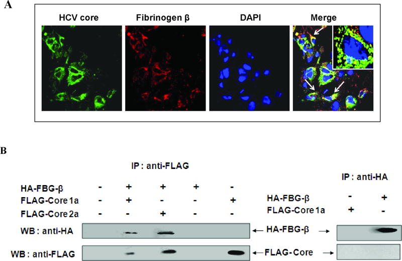
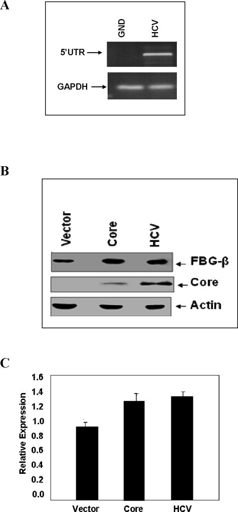
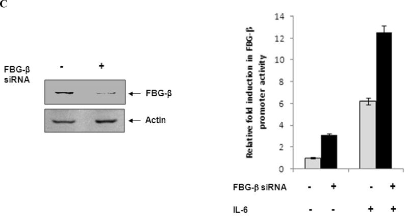
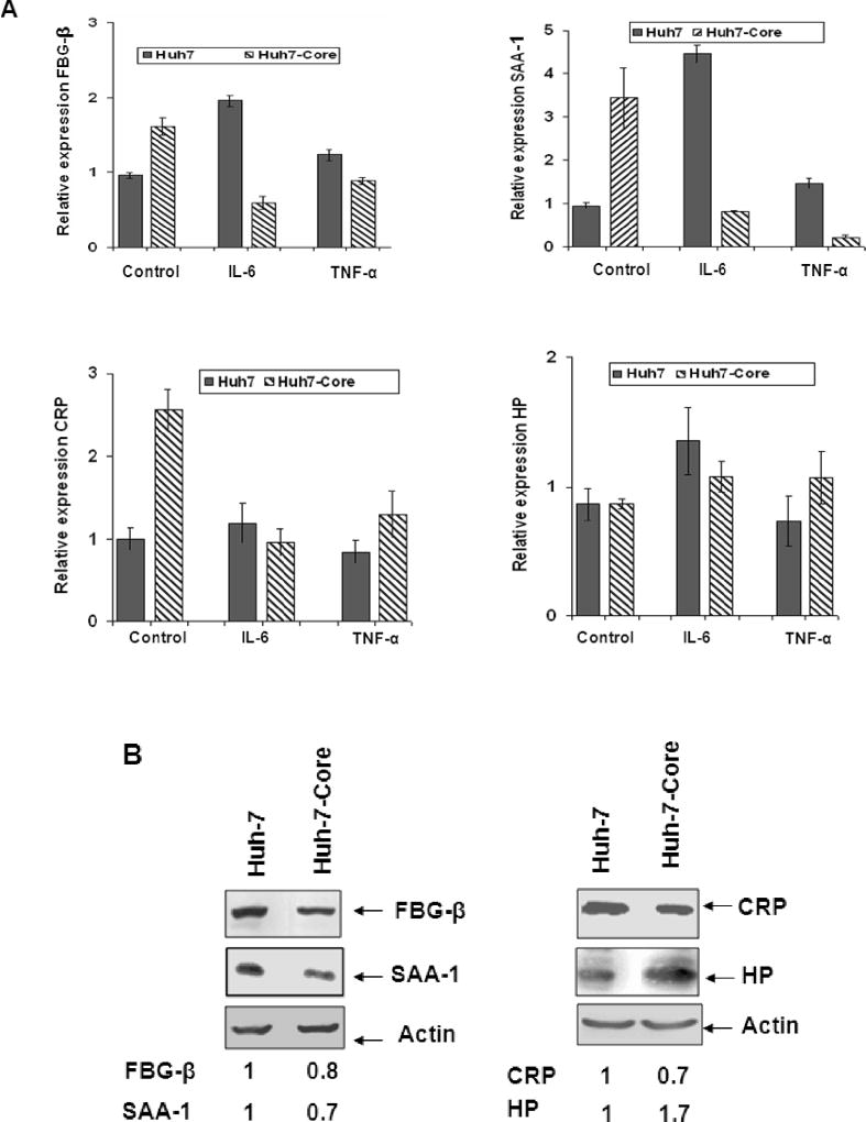
Fibrinogen-beta (FBG-beta), an important acute-phase protein (APP), is generated by the liver as a target for inflammatory mediators. Here we identified FBG-beta as a hepatitis C virus (HCV) core interacting protein by screening a human liver complementary DNA (cDNA) library using mammalian two-hybrid analysis. An association between FBG-beta and HCV core protein was verified by confocal microscopy and coimmunoprecipitation from the transfected human hepatocyte (Huh-7) cell line. HCV core or genomic RNA transfected Huh-7 cells modestly increased FBG-beta protein expression when compared to the basal level in control hepatocytes. Transfection of HCV core or full-length (FL) gene into Huh-7 cells up-regulated basal FBG-beta promoter activity. Exogenous addition of IL-6 stimulates FBG-beta promoter activity in hepatocytes. However, ectopic expression of HCV core or FL in hepatocytes inhibited IL-6-stimulated FBG-beta promoter activation. Inhibition of endogenous FBG-beta expression following introduction of small interfering RNA (siRNA) into cells displayed a gain of function of promoter regulation by HCV core protein. Further studies suggested that HCV core gene expression in stable transfectants of Huh-7 cells resulted in a basal up-regulation of FBG-beta and other APPs. However, treatment with cytokines, interleukin-6 (IL-6), or tumor necrosis factor-alpha repressed FBG-beta and other acute-phase response (APR) genes.

Conclusion: Our results reveal that the core/FBG-beta interaction may act as a regulatory feedback, allowing repression of IL-6-stimulated APR genes. Together, these data suggested a network of interactions between HCV core and the hepatic APR genes, and may contribute to impaired innate immunity for viral persistence.


Figures

Fig. 1
Fig. 1. Mammalian two-hybrid analysis for protein-protein interaction
Panel A: FACS analysis for hepatocellular protein interacting with HCV core by PCA. A human liver cDNA library was fused to fragment 1 of YFP (YFP[1]-cDNA library) and HCV core cDNA to fragment 2 (YFP2-core), in mammalian expression vectors harboring E. coli selection markers Ampicillin (Amp) and Kanamycin (Kan), respectively. COS-7 cells were cotransfected with core “bait” and human liver cDNA library “prey” fusions and positive clones indicating physical interaction between these proteins were identified by FACS. Panel B: A typical interaction of FBG-β protein with HCV core is shown from cotransfection of cells with the core and one of the identified clones (# 41) YFP fusions and detection by FACS. Dotted black line represents FBG-β clone and solid green line represent results from background control. Data were analyzed using FlowJo soft ware (Treestar). Percent of Max represents the number of events normalized according to FlowJo algorithm.
Fig. 2
Fig. 2. Association of fibrinogen-β and HCV core protein in hepatocytes
Panel A: Co-localization of FBG-β and HCV core protein by confocal microscopy. FBG-β gene was introduced into Huh-7-HCV core stable transfectants. Cells were stained with an antibody to core tagged with Alexa green, antibody to FBG-β tagged with Alexa red, and DAPI for nuclear staining. Co-localization of merged green and red colors is shown, and the arrows point out the merged yellow color in cells. The inset in the merged immunofluorescence image shows a higher magnification of a single cell. Panel B: Co-immunoprecipitation of FBG-β and HCV core proteins. Huh-7 cells were transfected with empty vector DNA (negative control), plasmid DNAs encoding HA-FBG-β and/or FLAG-core, indicated on top of each lane. Cell lysates were immunoprecipitated with an antibody to FLAG or HA, and probed with HA or FLAG specific antibody to detect HA-FBG-β or FLAG-core protein. The presence of HA-FBG-β in transfected cells is also shown after immunoprecipitation and detection by HA specific antibody.
Fig. 3
Fig. 3. HCV core protein expression modestly increases the basal level of FBG-β
Panel A: Full-length RNA from HCV genotype 1a transfected cells were separately examined after 6 days by RT-PCR for the presence viral RNA using 5’UTR primers. A polymerase defective HCV RNA (GND mutant) was similarly tested as a negative control. GAPDH gene expression was used to normalize HCV RNA expression. Panel B: Huh-7 cells (control), cells transfected with HCV core plasmid DNA, or electroporated with full-length RNA from genotype 1a were lysed and subjected to Western blot analysis using an antiserum to FBG-β or HCV core protein. The blot was reprobed with an antibody to actin to ascertain the level of protein load in each lane. Positions of the protein bands are indicated by arrows on the right. Panel C: The relative level of FBG-β from experiments in panel B was estimated by densitometric scanning after normalization against actin and shown as bar diagrams. Error bars represent standard errors from three independent experiments.
Fig. 4
Fig. 4. HCV core or FL transfected hepatocytes attenuate IL-6 stimulated FBG-β promoter activation
Panel A: Huh-7 cells were co-transfected with FBG-β-Luc (0.5 ug), and HCV core, FL or vector DNAs (250 ng). Luciferase activity was measured at 48 h post-transfection. Luciferase activities are presented as an average from three independent experiments with standard errors. The presence of HCV core protein in transfected cells were detected by Western blot analysis using a specific rabbit antiserum, and shown at the bottom of the bar diagram. Panel B: Transfected hepatocytes were treated with IL-6 (50 ng/ml) for 16 h before promoter assay. Fold difference of luciferase activity in the presence of IL-6 in HCV core or FL transfected hepatocytes in comparison to vector control is shown. Panel C: Huh-7 cells were electroporated with 10 µM siRNA to FBG-β in triplicate. Mock-electroporated cells were similarly used as a control. After 24 h, cells were co-transfected with FBG-β-Luc (0.5 ug), and HCV core (250 ng). Western blot analysis showing a reduction in endogenous FBG-β expression in transfected cells as compared to mock control is shown. Luciferase activity was measured at 48 h post-transfection. Transfected hepatocytes were treated with IL-6 (50 ng/ml) for 16 h before luciferase assay. Luciferase activities are presented as an average from three independent experiments with % errors.
Fig. 5
Fig. 5. Ectopic expression of HCV core protein attenuates cytokine stimulated acute phase response
Panel A: Real-time PCR analysis for mRNA expression of FBG-β, SAA-1, CRP, and HP was performed in control Huh-7 and HCV core stable transfectants of Huh-7 cells. Hepatocytes were treated with IL-6 (10 ng/ml) or TNF-α (20 ng/ml) for 6 h prior to RNA isolation. Standard error bars are shown from three different experiments. Panel B: Western blot analysis for FBG-β, SAA-1, CRP, and HP expression was performed using control Huh-7 and HCV core stable transfectants of Huh-7 cells. Hepatocytes were treated with IL-6 (10 ng/ml) for 6h prior to protein anlysis. The blots were run in two different sets (shown on left and right), and reprobed with an antibody to actin to ascertain the level of protein load in each lane. The relative level of specific proteins were estimated by densitometric scanning after normalization with actin, and the values are shown with that of control cells arbitrarily considered as 1 at the bottom.

Similar articles

Cited by

References

    1. Gabay C, Kushner I. Acute-phase proteins and other systemic responses to inflammation. N. Engl. J. Med. 1999;340:448–454. - PubMed
    1. Choy EH, Panayi GS. Cytokine pathways and joint inflammation in rheumatoid arthritis. N Engl J Med. 2001;344:907–16. - PubMed
    1. Baumann H, Gauldie J. The acute phase response. Immunol Today. 1994;15:74–80. - PubMed
    1. Black PH. The inflammatory response is an integral part of the stress response: Implications for atherosclerosis, insulin resistance, type II diabetes and metabolic syndrome X. Brain, Behav & Immun. 2003;17:350–64. - PubMed
    1. Gauldie J, Richards C, Harnish D, Lansdorp P, Baumann H. Interferon beta 2/B-cell stimulatory factor type 2 shares identity with monocyte-derived hepatocyte-stimulating factor and regulates the major acute phase protein response in liver cells. Proc. Natl. Acad. Sci. 1987;84:7251–7255. - PMC - PubMed

Publication types