

Induction of apoptosis by [8]-shogaol via reactive oxygen species generation, glutathione depletion, and caspase activation in human leukemia cells
- PMID: 20163181
- PMCID: PMC2990500
- DOI: 10.1021/jf904563c
Induction of apoptosis by [8]-shogaol via reactive oxygen species generation, glutathione depletion, and caspase activation in human leukemia cells
Abstract
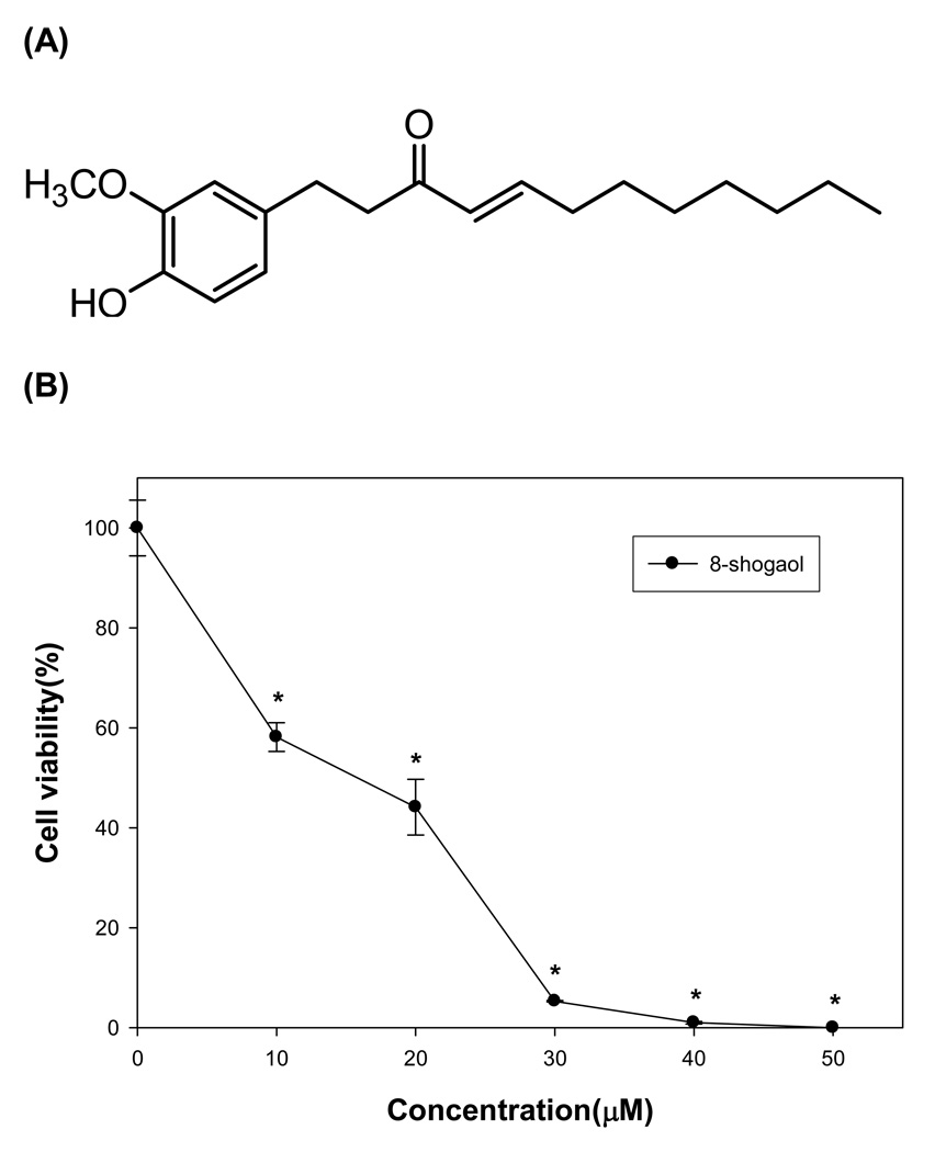
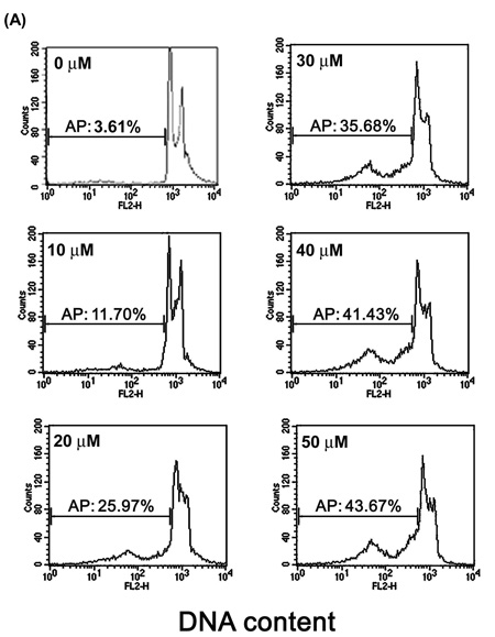
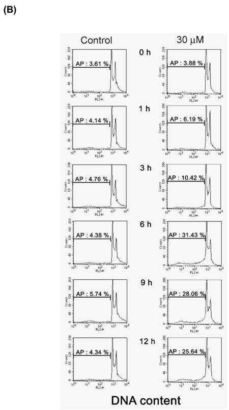
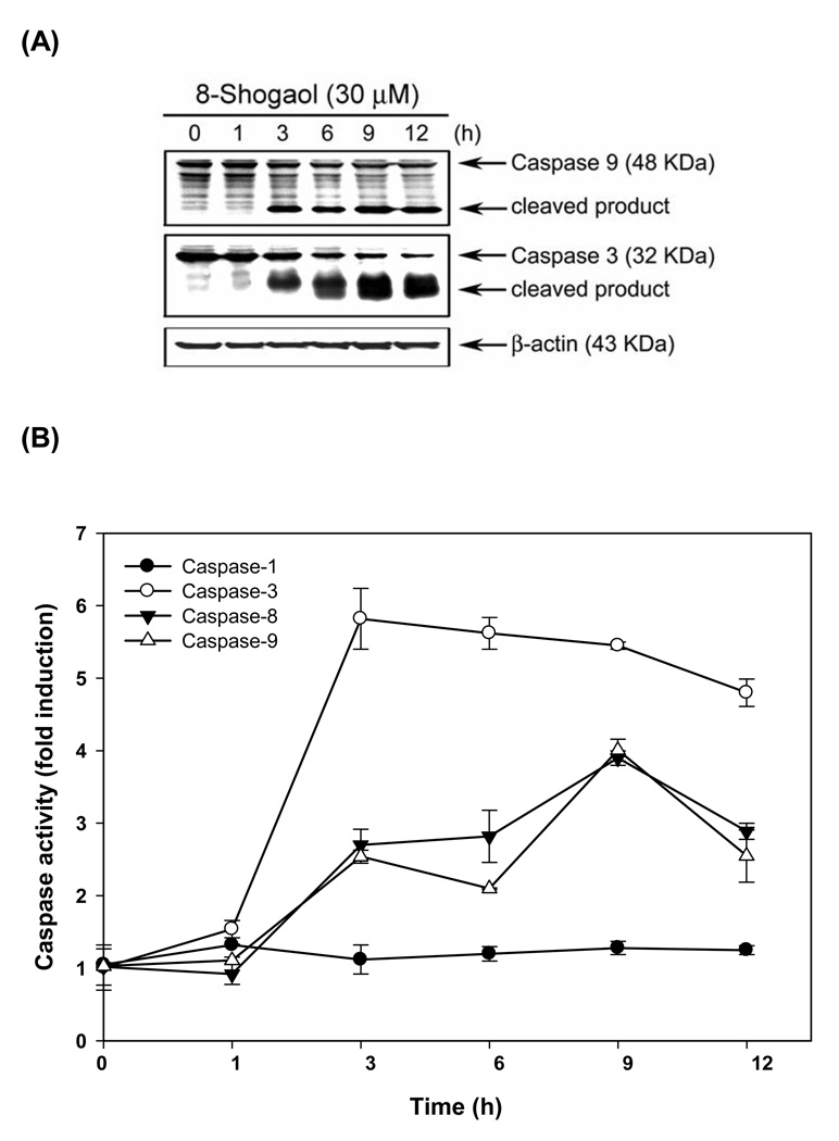
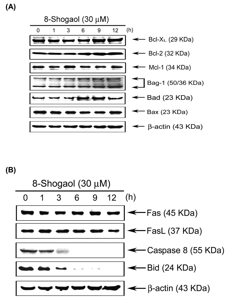
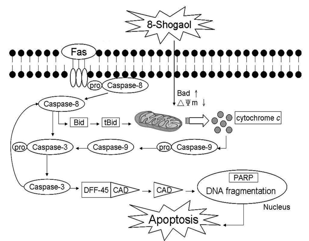
Ginger, the rhizome of Zingiber officinale , is a traditional medicine with a carminative effect and antinausea, anti-inflammatory, and anticarcinogenic properties. This study examined the growth inhibitory effects of [8]-shogaol, one of the pungent phenolic compounds in ginger, on human leukemia HL-60 cells. It demonstrated that [8]-shogaol was able to induce apoptosis in a time- and concentration-dependent manner. Treatment with [8]-shogaol caused a rapid loss of mitochondrial transmembrane potential, stimulation of reactive oxygen species (ROS) production, release of mitochondrial cytochrome c into cytosol, and subsequent induction of procaspase-9 and procaspase-3 processing. Taken together, these results suggest for the first time that ROS production and depletion of glutathione that contributed to [8]-shogaol-induced apoptosis in HL-60 cells.
Figures

Similar articles
-
6-Shogaol induces apoptosis in human colorectal carcinoma cells via ROS production, caspase activation, and GADD 153 expression.Mol Nutr Food Res. 2008 May;52(5):527-37. doi: 10.1002/mnfr.200700157. Mol Nutr Food Res. 2008. PMID: 18384088
-
Phytosome-Encapsulated 6-Gingerol- and 6-Shogaol-Enriched Extracts from Zingiber officinale Roscoe Protect Against Oxidative Stress-Induced Neurotoxicity.Molecules. 2024 Dec 22;29(24):6046. doi: 10.3390/molecules29246046. Molecules. 2024. PMID: 39770133 Free PMC article.
-
6-Shogaol enhances renal carcinoma Caki cells to TRAIL-induced apoptosis through reactive oxygen species-mediated cytochrome c release and down-regulation of c-FLIP(L) expression.Chem Biol Interact. 2015 Feb 25;228:69-78. doi: 10.1016/j.cbi.2015.01.020. Epub 2015 Jan 22. Chem Biol Interact. 2015. PMID: 25619640
-
An overview of 6-shogaol: new insights into its pharmacological properties and potential therapeutic activities.Food Funct. 2024 Jul 15;15(14):7252-7270. doi: 10.1039/d3fo04753a. Food Funct. 2024. PMID: 38287779 Review.
-
Occurrence, biological activity and metabolism of 6-shogaol.Food Funct. 2018 Mar 1;9(3):1310-1327. doi: 10.1039/c7fo01354j. Epub 2018 Feb 8. Food Funct. 2018. PMID: 29417118 Review.
Cited by
-
Natural Products and Acute Myeloid Leukemia: A Review Highlighting Mechanisms of Action.Nutrients. 2019 May 3;11(5):1010. doi: 10.3390/nu11051010. Nutrients. 2019. PMID: 31058874 Free PMC article. Review.
-
Herbal Formulation C168 Attenuates Proliferation and Induces Apoptosis in HCT 116 Human Colorectal Carcinoma Cells: Role of Oxidative Stress and DNA Damage.Evid Based Complement Alternat Med. 2016;2016:2091085. doi: 10.1155/2016/2091085. Epub 2016 Jan 17. Evid Based Complement Alternat Med. 2016. PMID: 26884792 Free PMC article.
-
6-Shogaol induces apoptosis in human hepatocellular carcinoma cells and exhibits anti-tumor activity in vivo through endoplasmic reticulum stress.PLoS One. 2012;7(6):e39664. doi: 10.1371/journal.pone.0039664. Epub 2012 Jun 29. PLoS One. 2012. PMID: 22768104 Free PMC article.
-
Inhibition of Polo-like kinase 1 (PLK1) triggers cell apoptosis via ROS-caused mitochondrial dysfunction in colorectal carcinoma.J Cancer Res Clin Oncol. 2023 Aug;149(10):6883-6899. doi: 10.1007/s00432-023-04624-2. Epub 2023 Feb 22. J Cancer Res Clin Oncol. 2023. PMID: 36810816 Free PMC article.
-
Elucidating the Pharmacological Properties of Zingiber officinale Roscoe (Ginger) on Muscle Ageing by Untargeted Metabolomic Profiling of Human Myoblasts.Nutrients. 2023 Oct 25;15(21):4520. doi: 10.3390/nu15214520. Nutrients. 2023. PMID: 37960173 Free PMC article.
References
-
- Sporn MB, Suh N. Chemoprevention of cancer. Carcinogenesis. 2000;21:525–530. - PubMed
-
- Aggarwal S, Ichikawa H, Takada Y, Sandur SK, Shishodia S, Aggarwal BB. Curcumin (diferuloylmethane) down-regulates expression of cell proliferation and antiapoptotic and metastatic gene products through suppression of IkappaBalpha kinase and Akt activation. Mol. Pharmacol. 2006;69:195–206. - PubMed
-
- Takada Y, Murakami A, Aggarwal BB. Zerumbone abolishes NF-kappaB and IkappaBalpha kinase activation leading to suppression of antiapoptotic and metastatic gene expression, upregulation of apoptosis, and downregulation of invasion. Oncogene. 2005;24:6957–6969. - PubMed
-
- Nishikawa T, Nakajima T, Moriguchi M, Jo M, Sekoguchi S, Ishii M, Takashima H, Katagishi T, Kimura H, Minami M, Itoh Y, Kagawa K, Okanoue T. A green tea polyphenol, epigalocatechin-3-gallate, induces apoptosis of human hepatocellular carcinoma, possibly through inhibition of Bcl-2 family proteins. J. Hepatol. 2006;44:1074–1082. - PubMed
-
- Park KK, Chun KS, Lee JM, Lee SS, Surh YJ. Inhibitory effects of [6]-gingerol, a major pungent principle of ginger, on phorbol ester-induced inflammation, epidermal ornithine decarboxylase activity and skin tumor promotion in ICR mice. Cancer Lett. 1998;129:139–144. - PubMed
Publication types
MeSH terms
Substances
Grants and funding
LinkOut - more resources
Full Text Sources
Medical
Research Materials
