Hydrazone ligation strategy to assemble multifunctional viral nanoparticles for cell imaging and tumor targeting
- PMID: 20163184
- PMCID: PMC3988696
- DOI: 10.1021/nl1002526
Hydrazone ligation strategy to assemble multifunctional viral nanoparticles for cell imaging and tumor targeting
Abstract
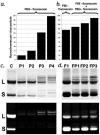
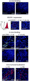
Multivalent nanoparticle platforms are attractive for biomedical applications because of their improved target specificity, sensitivity, and solubility. However, their controlled assembly remains a considerable challenge. An efficient hydrazone ligation chemistry was applied to the assembly of Cowpea mosaic virus (CPMV) nanoparticles with individually tunable levels of a VEGFR-1 ligand and a fluorescent PEGylated peptide. The nanoparticles recognized VEGFR-1 on endothelial cell lines and VEGFR1-expressing tumor xenografts in mice, validating targeted CPMV as a nanoparticle platform in vivo.
Figures


Similar articles
-
Development of viral nanoparticles for efficient intracellular delivery.Nanoscale. 2012 Jun 7;4(11):3567-76. doi: 10.1039/c2nr30366c. Epub 2012 Apr 16. Nanoscale. 2012. PMID: 22508503 Free PMC article.
-
CPMV-DOX delivers.Mol Pharm. 2013 Jan 7;10(1):3-10. doi: 10.1021/mp3002057. Epub 2012 Aug 6. Mol Pharm. 2013. PMID: 22827473 Free PMC article.
-
Interior engineering of a viral nanoparticle and its tumor homing properties.Biomacromolecules. 2012 Dec 10;13(12):3990-4001. doi: 10.1021/bm301278f. Epub 2012 Nov 14. Biomacromolecules. 2012. PMID: 23121655 Free PMC article.
-
Cowpea mosaic virus nanoparticles for cancer imaging and therapy.Adv Drug Deliv Rev. 2019 May;145:130-144. doi: 10.1016/j.addr.2019.04.005. Epub 2019 Apr 17. Adv Drug Deliv Rev. 2019. PMID: 31004625 Review.
-
Icosahedral plant viral nanoparticles - bioinspired synthesis of nanomaterials/nanostructures.Adv Colloid Interface Sci. 2017 Oct;248:1-19. doi: 10.1016/j.cis.2017.08.005. Epub 2017 Aug 31. Adv Colloid Interface Sci. 2017. PMID: 28916111 Review.
Cited by
-
Development of viral nanoparticles for efficient intracellular delivery.Nanoscale. 2012 Jun 7;4(11):3567-76. doi: 10.1039/c2nr30366c. Epub 2012 Apr 16. Nanoscale. 2012. PMID: 22508503 Free PMC article.
-
Radiolabeling and evaluation of (64)Cu-DOTA-F56 peptide targeting vascular endothelial growth factor receptor 1 in the molecular imaging of gastric cancer.Am J Cancer Res. 2015 Oct 15;5(11):3301-10. eCollection 2015. Am J Cancer Res. 2015. PMID: 26807312 Free PMC article.
-
The art of engineering viral nanoparticles.Mol Pharm. 2011 Feb 7;8(1):29-43. doi: 10.1021/mp100225y. Epub 2010 Dec 17. Mol Pharm. 2011. PMID: 21047140 Free PMC article. Review.
-
Design of virus-based nanomaterials for medicine, biotechnology, and energy.Chem Soc Rev. 2016 Jul 25;45(15):4074-126. doi: 10.1039/c5cs00287g. Chem Soc Rev. 2016. PMID: 27152673 Free PMC article. Review.
-
Nanocaged platforms: modification, drug delivery and nanotoxicity. Opening synthetic cages to release the tiger.Nanoscale. 2017 Jan 26;9(4):1356-1392. doi: 10.1039/c6nr07315h. Nanoscale. 2017. PMID: 28067384 Free PMC article. Review.
References
-
- Ntziachristos V, Ripoll J, Wang LV, Weissleder R. Nat Biotechnol. 2005;23(3):313–20. - PubMed
-
- Wang Q, Lin T, Johnson JE, Finn MG. Chem Biol. 2002;9(7):813–9. - PubMed
-
- Lin T, Chen Z, Usha R, Stauffacher CV, Dai JB, Schmidt T, Johnson JE. Virology. 1999;265(1):20–34. - PubMed
-
- Chatterji A, Ochoa W, Paine M, Ratna BR, Johnson JE, Lin T. Chem Biol. 2004;11(6):855–63. - PubMed
Publication types
MeSH terms
Substances
Grants and funding
LinkOut - more resources
Full Text Sources
Other Literature Sources

