

Extrahippocampal gray matter loss and hippocampal deafferentation in patients with temporal lobe epilepsy
- PMID: 20163442
- PMCID: PMC2855766
- DOI: 10.1111/j.1528-1167.2009.02506.x
Extrahippocampal gray matter loss and hippocampal deafferentation in patients with temporal lobe epilepsy
Abstract
Purpose: Medial temporal epilepsy (MTLE) is associated with extrahippocampal brain atrophy. The mechanisms underlying brain damage in MTLE are unknown. Seizures may lead to neuronal damage, but another possible explanation is deafferentation from loss of hippocampal connections. This study aimed to investigate the relationship between hippocampal deafferentation and brain atrophy in MTLE.
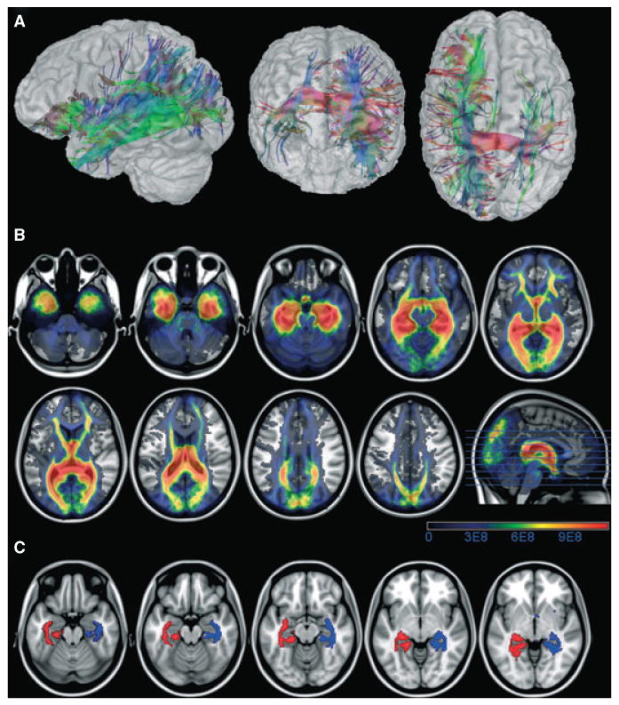
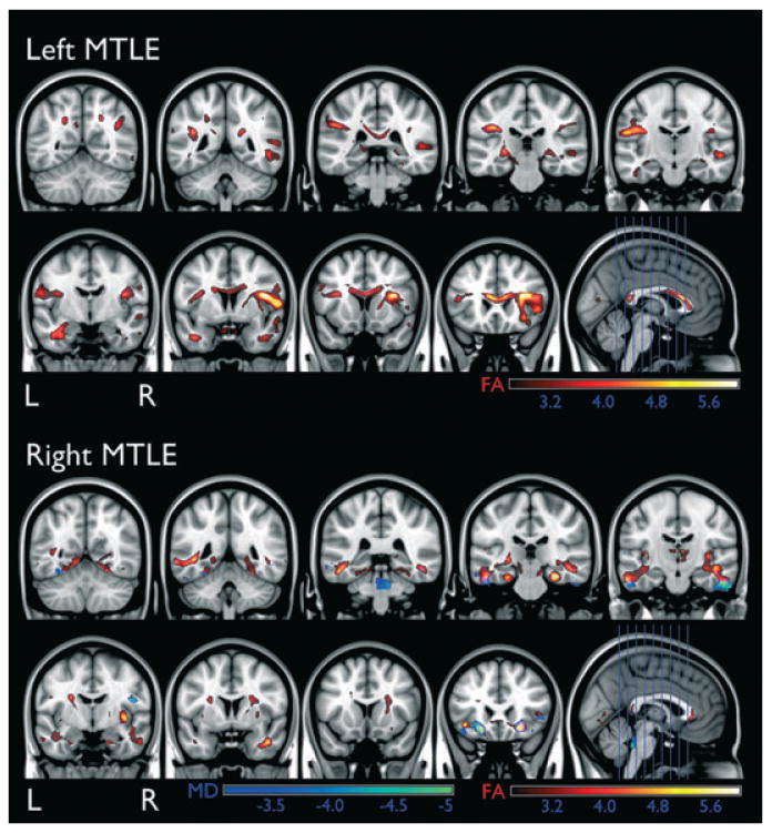
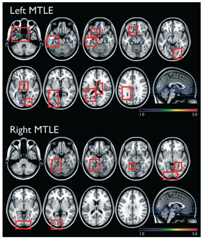
Methods: Three different MRI studies were performed involving 23 patients with unilateral MTLE (8 left and 15 right) and 34 healthy controls: (1) voxel-based morphometry (VBM), (2) diffusion tensor imaging (DTI) and (3) probabilistic tractography (PT). VBM was employed to define differences in regional gray matter volume (GMV) between controls and patients. Voxel-wise analyses of DTI evaluated differences in fractional anisotropy (FA), mean diffusivity (MD) and hippocampal PT. Z-scores were computed for regions-of-interest (ROI) GMV and peri-hippocampal FA and MD (to quantify hippocampal fiber integrity). The relationship between hippocampal deafferentation and regional GMV was investigated through the association between ROI Z scores and hippocampal fiber integrity.
Results: Patients with MTLE exhibited a significant reduction in GMV and FA in perihippocampal and limbic areas. There was a decrease in hippocampal PT in patients with MTLE in limbic areas. A significant relationship between loss of hippocampal connections and regional GMV atrophy was found involving the putamen, pallidum, middle and inferior temporal areas, amygdala and ceberellar hemisphere.
Discussion: There is a relationship between hippocampal disconnection and regional brain atrophy in MTLE. These results indicate that hippocampal deafferentation plays a contributory role in extrahippocampal brain damage in MTLE.
Conflict of interest statement
Figures
References
-
- Alessio A, Bonilha L, Rorden C, Kobayashi E, Min LL, Damasceno BP, Cendes F. Memory and language impairments and their relationships to hippocampal and perirhinal cortex damage in patients with medial temporal lobe epilepsy. Epilepsy Behav. 2006;8:593–600. - PubMed
-
- Ashburner J, Friston KJ. Unified segmentation. Neuroimage. 2005;26:839–851. - PubMed
-
- Bernasconi N, Bernasconi A, Caramanos Z, Antel SB, Andermann F, Arnold DL. Mesial temporal damage in temporal lobe epilepsy: a volumetric MRI study of the hippocampus, amygdala and parahippocampal region. Brain. 2003;126:462–469. - PubMed
-
- Bernasconi N, Duchesne S, Janke A, Lerch J, Collins DL, Bernasconi A. Whole-brain voxel-based statistical analysis of gray matter and white matter in temporal lobe epilepsy. Neuroimage. 2004;23:717–723. - PubMed
MeSH terms
Grants and funding
LinkOut - more resources
Full Text Sources
Medical
