. 2010 Feb;17(2):113-27.
doi: 10.1111/j.1549-8719.2009.00011.x.

Modulation of microvascular smooth muscle adhesion and mechanotransduction by integrin-linked kinase

Affiliations

Modulation of microvascular smooth muscle adhesion and mechanotransduction by integrin-linked kinase

Shaoxing Huang et al. Microcirculation. 2010 Feb.

Abstract

Objective: In this study, we investigated the involvement of integrin-linked kinase (ILK) in the adhesion of arteriolar vascular smooth muscle cells (VSMC) to fibronectin (FN) and in the mechano-responsiveness of VSMC focal adhesions (FA).

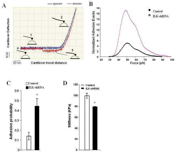
Methods: ILK was visualized in VSMC by expressing EGFP-ILK and it was knocked down using ILK-shRNA constructs. Atomic force microscopy (AFM) was used to characterize VSMC interactions with FN, VSMC stiffness and to apply and measure forces at a VSMC single FA site.

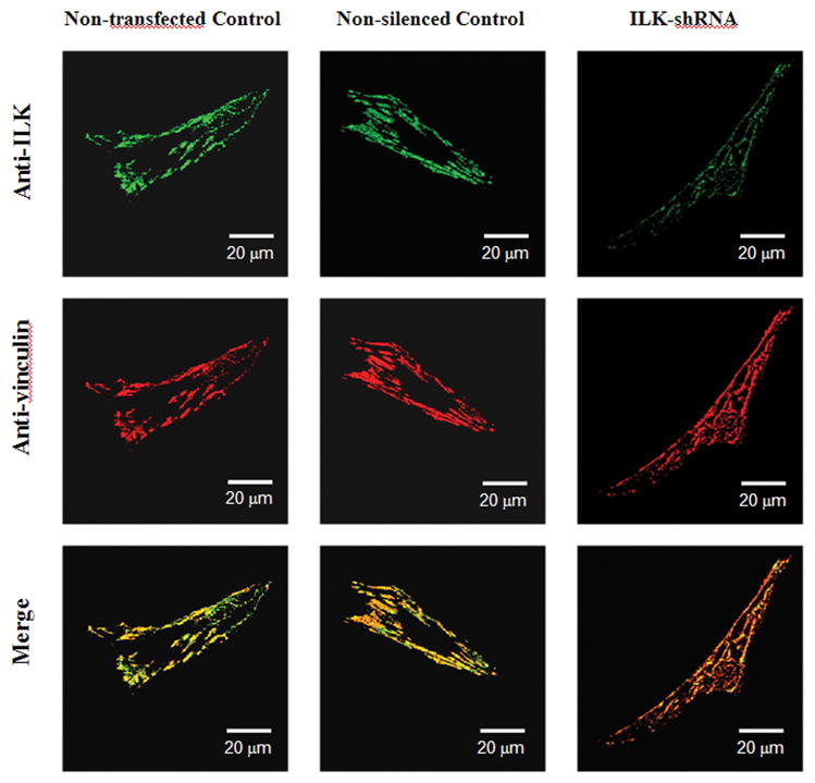
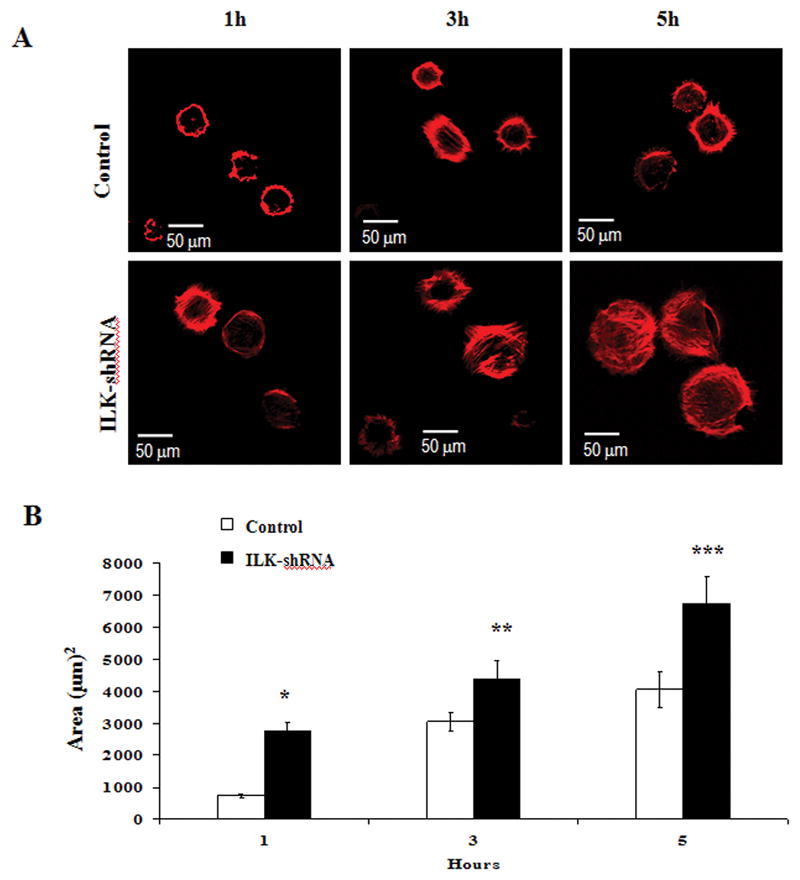
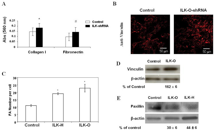
Results: ILK was localized to FA and silencing ILK promoted cell spreading, enhanced cell adhesion, reduced cell proliferation and reduced downstream phosphorylation of GSK-3beta and PKB/Akt. AFM studies demonstrated that silencing ILK enhanced alpha5beta1 integrin adhesion to FN and enhanced VSMC contraction in response to a pulling force applied at the level of a single FN-FA site.

Conclusions: ILK functions in arteriolar VSMC appear linked to multiple signaling pathways and processes that inhibit cell spreading, cell adhesion, FA formation, adhesion to FN and the mechano-responsiveness of FN-FA sites.


Figures

Figure 1
Figure 1
Confocal fluorescent images of cultured vascular smooth muscle cells transfected with EGFP-ILK (A) or EGFP alone (B). EGFP-ILK was localized to focal adhesion sites (arrows). In comparison, EGFP alone was diffusely dispersed throughout the cell. (C) Immunofluorescent labeling showed the presence of ILK (green) in the vascular smooth muscle of an intact, isolated and pressurized rat cremaster feed arteriole. The ILK is apparent as punctate labeling on the cell surface as well as some diffuse labeling in the cytosol. The cell nuclei were shown in red. Scale bar = 10 μm.
Figure 2
Figure 2
Immunofluorescent images of vascular smooth muscle cells probed with anti-ILK and anti-vinculin antibodies showed that ILK colocalized with focal adhesion protein vinculin at focal adhesions. Results are representative of three independent experiments. Scale bar = 30 μm. Colocalization index was calculated with Olympus FV1000 software and was presented as a mean ± SE of 10 cells. Non-transfected control, cells were cultured less than 10 passages and not transfected (Colocalization index, 0.56 ± 0.02); Non-silenced control cells, cells were cultured between 10 and 20 passages and transfected with non-silenced control construct (Colocalization index, 0.73 ± 0.01); ILK-shRNA, cells were cultured between 10 and 20 passages and transfected with ILK-shRNA silencing construct (Colocalization index, 0.69 ± 0.02). As shown, ILK localizations in VSMC were not affected by transfection with either non-silenced or ILK-shRNA constructs.
Figure 3
Figure 3
(A) and (B) Western blots and immunofluorescence probed with an anti-ILK antibody showed that ILK expression was knocked down in VSMC stably transfected with ILK-O and ILK-H construct compared to cells transfected with non-silenced control (A control of non-transfected cells: WT-control, is also included in Western blotting). Images were taken with a 40 × 1.30 objective. Scale bar = 50 μm. Blots were quantified and normalized to β-actin and displayed as percent of control. (C) and (D): ILK silencing reduces phosphorylation of GSK-3β and AKT1/PKB in VSMC stably transfected with ILK-O construct compared to cells transfected with non-silenced control. Western blots were probed with antibodies against phospho-specific GSK-3β (P-GSK-3β), total GSK-3β (T-GSK-3β), phospho-specific AKT1/PKB (P-AKT1/PKB) and total AKT1/PKB(T-AKT1/PKB). Blots were quantified and normalized to total GSK-3β or total AKT1/PKB and displayed as percent of the control. The results are representative of 3 independent experiments.
Figure 4
Figure 4. ILK silencing promotes VSMC spreading
(A) VSMC stably transfected with non-silenced control and ILK-shRNA constructs were plated with serum-free culture medium on 50 μg/ml fibronectin-coated dishes for 1, 3 and 5h and were then fixed and stained with Alexa 568-conjugated-phalloidin. (B) Bar graphs representing mean cell areas (in square micrometers) calculated using Image J. (*, p<0.001 vs. control; **, p<0.01 vs. control; ***, p<0.05 vs. control). Error bars represent standard error of the mean. ILK silencing increases cell spreading in ILK-shRNA cells in comparison with non-silencing control. Images were taken with a 40 × 1.30 objective. Results are representative of 4 independent experiments. Scale bar = 50 μm.
Figure 5
Figure 5. ILK silencing inhibits cell migration and proliferation of VSMC
(A) Migration assays were carried out in Boyden migration chamber with VSMCs transfected with non-silenced control and ILK-shRNA constructs (ILK-O and ILK-H). The stimulants in lower chambers are as follows: null: un-stimulated control; PDGF: PDGF, 2.5 μg/ml; FN: 50μg/ml fibronectin. Data are displayed as an index relative to the control in equivalent condition. Results are based on at least 3 independent experiments performed at least in quadruplicate. (B) Proliferation assays of control and ILK-O transfected VSMC using cell counting. Error bars represent the standard error between experiments. * p<0.05, n=3; **p<0.01, n=5.
Figure 6
Figure 6. ILK silencing enhances cell adhesion in VSMC
(A) Control or ILK-shRNA constructs stably transfected VSMC were plated in 96-well plates coated with 50 μg/ml collagen type I or 50μg/ml FN and allowed to adhere for 1 h. Results represent mean ± SE of 6 independent experiments. (*, p<0.05 vs. control; #, p<0.05 vs. control). (B) VSMC stably transfected with non-silened control and ILK-shRNA construct (ILK-O) were plated with serum-free culture medium on 50 μg/ml FN-coated dishes and immunofluorescence was probed with anti-vinculin antibody. Images were taken with a 40 × 1.30 objective. Scale bar = 50 μm. Results are representative of 4 independent experiments. (C) Focal adhesion number was quantified using NIH Image J as described in Materials and Methods. Values are mean of focal adhesions per cell. (n=22, p<0.01 vs. control). (D) Western blots showed that vinculin expression is increased in ILK-O VSMC in comparison with non-silenced control (162 ± 6% of control). (E) paxillin expression is decreased in ILK-O and ILK-H VSMC in comparison with non-silenced control (162 ± 6% of control). Blots were quantified and normalized to actin and displayed as percent of the control. Results are representative of 3 independent experiments, p<0.05 for both vinculin and paxillin blotts.
Figure 7
Figure 7
(A) Western blot showing that Hic-5 Expression was enhanced in ILK-O and ILK-H comparing to control. (B) Immunofluorescece confocal images of paxillin and Hic-5 at VSMC focal adhesions. Paxillin was labeled with K-20 antibody, Alexa-647; and Hic-5 was labeled with 34/Hic-5, Alexa-488. Both paxillin and hic-5 were localized at focal adhesions in either control or ILK knock-down VSMCs. Similar results were observed in ILK-O VSMCs (data not shown).
Figure 8
Figure 8
(A) A typical AFM recorded force curve. The probe was coated with FN, and an adhesion event is detected by the “snap-off” in the retraction curve. The compression stiffness was measured by fitting the approaching curve with Hertz model. (B) The adhesion forces (unbinding or rupture forces) distribution plotted as a histogram. (C) The probability of binding was determined as (number of force curves with adhesions)/(total number of force curves). Error bars represent standard error of the mean. Compared to control VSMC, ILK-shRNA cells exhibited an increased probability of binding to fibronectin but the adhesion force distribution was not significantly different. (D) Stiffness of control and ILK-O cells. Error bars represent standard error of the mean. *: p<0.05.
Figure 9
Figure 9. ILK silencing enhanced pulling force response in VSMC
(A) The AFM cantilever with FN-coated bead was brought into contact with the cell surface of VSMC (control or ILK-O). The bead was then pulled from the cell surface using the AFM in the Z-axis, and the bead displacement over 250 seconds was shown to indicate the VSMC response to the pulling force. Error bars represent standard error of the mean. (n=5, p<0.05 vs. control). (B) Percentage of cell compensation as an indicator of quantified cellular micro-myogenic response. (C) Apparent pulling elasticity as an indicator of the strength of the focal adhesion and the associated cytoskeleton underneath. Error bars represent standard error of the mean.

Similar articles

Cited by

References

    1. Adams JC. Cell-matrix contact structures. Cell Mol Life Sci. 2001;58(3):371–92. - PMC - PubMed
    1. Attwell S, Mills J, et al. Integration of cell attachment, cytoskeletal localization, and signaling by integrin-linked kinase (ILK), CH-ILKBP, and the tumor suppressor PTEN. Mol Biol Cell. 2003;14(12):4813–25. - PMC - PubMed
    1. Brakebusch C, Fassler R. The integrin-actin connection, an eternal love affair. EMBO J. 2003;22(10):2324–33. - PMC - PubMed
    1. Brummelkamp TR, Bernards R, et al. A system for stable expression of short interfering RNAs in mammalian cells. Science. 2002;296(5567):550–3. - PubMed
    1. Chao JT, Martinez-Lemus LA, et al. Modulation of alpha7-integrin-mediated adhesion and expression by platelet-derived growth factor in vascular smooth muscle cells. Am J Physiol Cell Physiol. 2006;290(4):C972–80. - PubMed

Publication types

MeSH terms