. 2010 May;11(5):587-600.
doi: 10.1111/j.1600-0854.2010.01050.x. Epub 2010 Feb 15.

Legionella pneumophila promotes functional interactions between plasma membrane syntaxins and Sec22b

Affiliations

Legionella pneumophila promotes functional interactions between plasma membrane syntaxins and Sec22b

Kohei Arasaki et al. Traffic. 2010 May.

Abstract

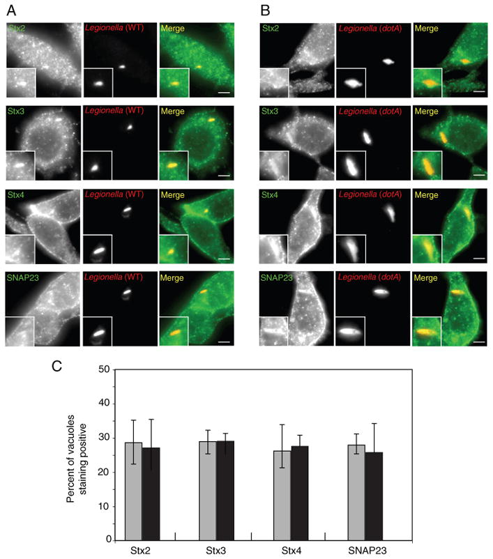
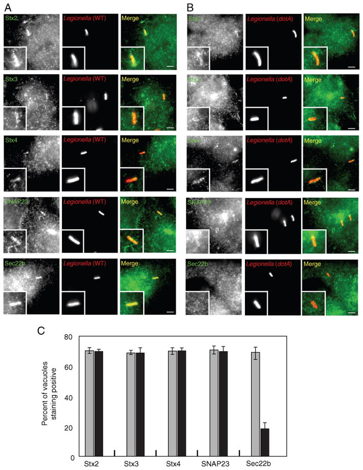
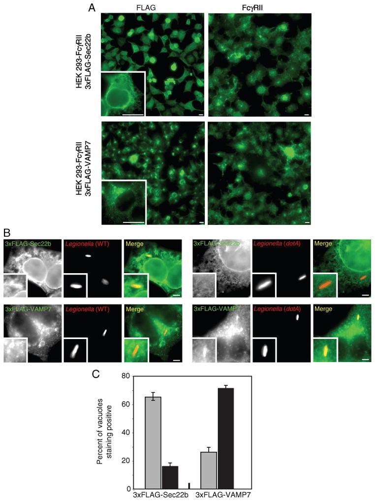
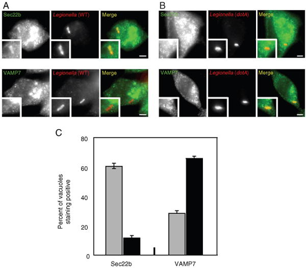
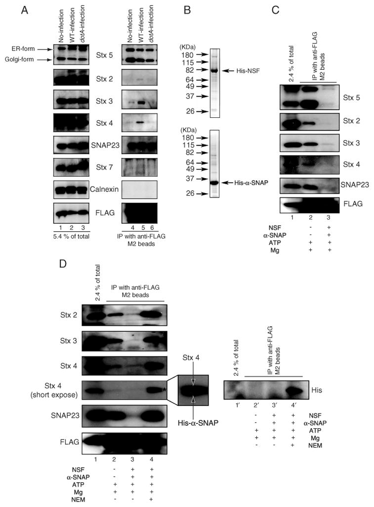
Biogenesis of a specialized organelle that supports intracellular replication of Legionella pneumophila involves the fusion of secretory vesicles exiting the endoplasmic reticulum (ER) with phagosomes containing this bacterial pathogen. Here, we investigated host plasma membrane SNARE proteins to determine whether they play a role in trafficking of vacuoles containing L. pneumophila. Depletion of plasma membrane syntaxins by RNA interference resulted in delayed acquisition of the resident ER protein calnexin and enhanced retention of Rab1 on phagosomes containing virulent L. pneumophila, suggesting that these SNARE proteins are involved in vacuole biogenesis. Plasma membrane-localized SNARE proteins syntaxin 2, syntaxin 3, syntaxin 4 and SNAP23 localized to vacuoles containing L. pneumophila. The ER-localized SNARE protein Sec22b was found to interact with plasma membrane SNAREs on vacuoles containing virulent L. pneumophila, but not on vacuoles containing avirulent mutants of L. pneumophila. The addition of alpha-SNAP and N-ethylmaleimide-sensitive factor (NSF) to the plasma membrane SNARE complexes formed by virulent L. pneumophila resulted in the dissociation of Sec22b, indicating functional pairing between these SNAREs. Thus, L. pneumophila stimulates the non-canonical pairing of plasma membrane t-SNAREs with the v-SNARE Sec22b to promote fusion of the phagosome with ER-derived vesicles. The mechanism by which L. pneumophila promotes pairing of plasma membrane syntaxins and Sec22b could provide unique insight into how the secretory vesicles could provide an additional membrane reserve subverted during phagosome maturation.


Figures

Figure 1
Figure 1
Dynamics of Sec22b, calnexin and Rab1b recruitment to vacuoles containing L. pneumophila after silencing of plasma membrane syntaxins. A) Cell lysates were obtained from HEK293-FcγRII cells transfected for 72 h with the siRNA indicated (top), and immunoblot analysis was used to measure the levels of the proteins indicated (left). Calnexin levels were used to assess equal protein loading. B) HEK293-FcγRII cells were treated with the indicated siRNA or mock treated as indicated in the upper left corner of each image panel. Cells were fixed and stained with antibodies specific for the proteins indicated on the left of each row. FcγRII staining was conducted to determine if the triple syntaxin knockdown (Stx2/3/4 KD) affected the localization of this plasma membrane protein. Scale bar = 5 μm. C and D) HEK293-FcγRII cells were transfected with siRNA targeting the indicated syntaxins (Stx2, Stx3, Stx4, Stx2/3/4) or treated with control siRNA (mock) as indicated at the bottom of the graph. Cells were fixed either 1 (C) or 4 h (D) after infection with wild-type L. pneumophila, and stained with antibodies to either Sec22b, calnexin or Rab1b as indicated in each panel. Colocalization of Sec22b, calnexin and Rab1b was assessed by fluorescence microscopy and the data are presented as the percent of vacuoles that were positive for each marker. Data are the mean ± SEM of three independent experiments in which 100 vacuoles were scored [*p < 0.02 and **p < 0.04 (C), *p < 0.002 and **p < 0.001 (D), as compared with mock-treated]. E) HEK293-FcγRII cells were transfected with an siRNA pool to silence the production of the plasma membrane-localized syntaxins (Stx2/3/4; black bars) or treated with control siRNA (mock; white bars) as indicated. Cells were fixed 8 h after infection with wild-type L. pneumophila. Intracellular replication of L. pneumophila was assessed by counting the number of bacteria residing in a single vacuole in the infected cells. The data are presented as the percent of vacuoles in mock-transfected cells (white bars) and Stx2/3/4 siRNA-transfected cells (black bars) that contained 1–3 bacteria, 4–6 bacteria or >7 bacteria. Data are the mean ± SEM of three independent experiments in which 100–120 vacuoles were scored (*p < 0.009 and **p < 0.02, as compared with mock-treated).
Figure 2
Figure 2
Localization of endogenous plasma membrane SNAREs to vacuoles containing L. pneumophila in RAW 264.7 cells. A and B) RAW 264.7 macrophage-like cells were infected with dsRed-expressing wild-type L. pneumophila (A) or the dotA mutant (B). At 1 h after infection, the cells were fixed and stained with antibodies specific for the proteins listed on the left (Stx2, Stx3, Stx4 or SNAP23). The insert contains a magnified image of the vacuole from each panel. Bar = 1 μm. C) Localization of the SNAREs indicated at the bottom of the graph (Stx2, Stx3, Stx4, SNAP23) was assessed for vacuoles containing either wild-type L. pneumophila (gray bars) or a dotA mutant (black bars) at 1 h after infection. Values are the mean ± SEM of three independent experiments in which 70 vacuoles were scored.
Figure 3
Figure 3
Localization of endogenous plasma membrane SNAREs to vacuoles containing L. pneumophila in HEK293-FcγRII cells. A and B) HEK293-FcγRII cells were infected with dsRed-expressing wild-type L. pneumophila (A) or the dotA mutant (B). At 1 h after infection, the cells were fixed and stained with antibodies specific for the protein listed on the left (Stx2, Stx3, Stx4, SNAP23 or Sec22b). The insert contains a magnified image of the vacuole from each panel. Bar = 1 μm. C) Localization of the protein indicated at the bottom of the graph (Stx2, Stx3, Stx4, SNAP23, Sec22b) was assessed for vacuoles containing either wild-type L. pneumophila (gray bars) or a dotA mutant (black bars) at 1 h after infection. Values are the mean ± SEM of three independent experiments in which 70 vacuoles were scored.
Figure 4
Figure 4
Differential localization of FLAG-tagged VAMP7 and Sec22b to vacuoles containing L. pneumophila in HEK293-FcγRII cells. A) Fluorescence micrographs of stable HEK293-FcγRII cell lines producing either 3x-FLAG-Sec22b (top panels) or 3x-FLAG-VAMP7 (bottom panels). Cells were stained with an antibody specific for FLAG (left panels) or an antibody specific for FcγRII (right panels). A higher magnification of a representative cell stained with the anti-FLAG antibody is shown in the insert. Bar = 5 μm. B) Fluorescence micrographs of stable HEK293-FcγRII cell lines producing either 3x-FLAG-Sec22b (top panels) or 3x-FLAG-VAMP7 (bottom panels) with either wild-type L. pneumophila (left series) or a dotA mutant (right series) at 1 h after infection. Anti-FLAG staining is shown in green and the L. pneumophila staining is shown in red in the merged panels. The insert contains a magnified image of the vacuole from each panel. Bar = 1 μm. C) Localization of 3x-FLAG-Sec22b and 3x-FLAG-VAMP7 was assessed for vacuoles containing either wild-type L. pneumophila (gray bars) or a dotA mutant (black bars) at 1 h after infection. Values are the mean ± SEM of three independent experiments in which 100 vacuoles were scored.
Figure 5
Figure 5
Differential localization of endogenous VAMP7 and Sec22b to vacuoles containing L. pneumophila in RAW 264.7 cells. A and B) Fluorescence micrographs of RAW 264.7 macrophage-like cells stained with an antibody specific for Sec22b (top panels) or VAMP7 (bottom panels) with either wild-type L. pneumophila (A) or a dotA mutant (B) at 1 h after infection. The insert contains a magnified image of the vacuole from each panel. Bar = 1 μm. C) Localization of Sec22b and VAMP7 was assessed for vacuoles containing either wild-type L. pneumophila (gray bars) or a dotA mutant (black bars) at 1 h after infection. Values are the mean ± SEM of three independent experiments in which 100 vacuoles were scored.
Figure 6
Figure 6
Sec22b forms a complex with plasma membrane t-SNAREs upon infection with wild-type L. pneumophila. A) HEK293-FcγRII 3x-FLAG-Sec22b cells were infected without (lanes 1 and 4) or with wild-type L. pneumophila (lanes 2 and 5) or dotA mutant (lanes 3 and 6). Sec22b was immunoprecipitated with anti-FLAG M2 beads from lysates prepared 1 h after infection. A portion of the cell lysate (5.4% of total; left lanes) and the immunoprecipitated proteins (IP with anti-FLAG M2 beads; right lanes) were separated by SDS PAGE and the amounts of the indicated proteins (Stx5, Stx2, Stx3, Stx4, SNAP23, calnexin, FLAG-Sec22b) were determined by immunoblot analysis. B) Recombinant His6-NSF and His6-α-SNAP were separated by SDS PAGE and stained with coomasie blue. Indicated on the left are the positions of weight markers. C) HEK293-FcγRII 3x-FLAG-Sec22b cells were infected with wild-type L. pneumophila. Sec22b was immunoprecipitated with anti-FLAG M2 beads from lysates prepared 1 h after infection. As indicated in the bottom legend, cell lysates were treated with 0.5 mm ATP and 8 mm MgCl2 (lane 2) or 0.5 mm ATP, 8 mm MgCl2, 10 μg/mL His6-NSF and 5 μg/mL His6-α-SNAP (lane 3) at 16°C for 1 h prior to immunoprecipitation. A portion of the cell lysate (2.4% of total; lane 1) and the immunoprecipitated proteins (IP with anti-FLAG M2 beads; lanes 2 and 3) were separated by SDS PAGE and the amounts of the indicated proteins (Stx5, Stx2, Stx3, Stx4, SNAP23, FLAG-Sec22b) were determined by immunoblot analysis. D) HEK293-FcγRII 3x-FLAG-Sec22b cells were infected with wild-type L. pneumophila. Sec22b was immunoprecipitated with anti-FLAG M2 beads from lysates prepared 1h after infection. As indicated in the bottom legend, cell lysates were treated with 0.5 mm ATP and 8 mm MgCl2(lanes 2 and 2′ ), 0.5 mm ATP, 8 mm MgCl2, 10 μg/mL His6-NSF and 5 μg/mL His6-α-SNAP (lanes 3 and 3′) or 0.5 mm ATP, 8 mm MgCl2, 10 μg/mL NEM-pretreated His6-NSF and 5 μg/mL His6-α-SNAP (lanes 4 and 4′) at 16°C for 1 h. After incubation, the samples were immunoprecipitated with anti-FLAG M2 beads. A portion of the cell lysate (2.4% of total; lanes 1 and 1′) and the immunoprecipitated proteins (IP with anti-FLAG M2 beads) were separated by SDS PAGE and the amounts of the indicated proteins (Stx2, Stx3, Stx4, SNAP23, FLAG-Sec22b) were determined by immunoblot analysis in lanes 1–4. Asterisk and magnification of the Stx4 band in lane 4 shows the presence of His6-α-SNAP, which was detected by the anti-Stx4 antibody. An anti-His immunoblot shown on the right (lanes 1′ – 4′) confirms His6-α-SNAP was immunoprecipitated with FLAG-Sec22b in the NEM-treated NSF-containing sample (lane 4′).

Similar articles

Cited by

References

    1. Kumar Y, Valdivia RH. Leading a sheltered life: intracellular pathogens and maintenance of vacuolar compartments. Cell Host Microbe. 2009;5:593–601. - PMC - PubMed
    1. Swanson MS, Isberg RR. Association of Legionella pneumophila with the macrophage endoplasmic reticulum. Infect Immun. 1995;63:3609–3620. - PMC - PubMed
    1. Kagan JC, Roy CR. Legionella phagosomes intercept vesicular traffic from endoplasmic reticulum exit sites. Nat Cell Biol. 2002;4:945–954. - PubMed
    1. Derre I, Isberg RR. Legionella pneumophila replication vacuole formation involves rapid recruitment of proteins of the early secretory system. Infect Immun. 2004;72:3048–3053. - PMC - PubMed
    1. Kagan JC, Stein MP, Pypaert M, Roy CR. Legionella subvert the functions of Rab1 and Sec22b to create a replicative organelle. J Exp Med. 2004;199:1201–1211. - PMC - PubMed

Publication types

MeSH terms

LinkOut - more resources