. 2010 Mar-Apr;18(2):193-201.
doi: 10.1111/j.1524-475X.2010.00570.x. Epub 2010 Feb 16.

Impaired angiogenesis and mobilization of circulating angiogenic cells in HIF-1alpha heterozygous-null mice after burn wounding

Affiliations

Impaired angiogenesis and mobilization of circulating angiogenic cells in HIF-1alpha heterozygous-null mice after burn wounding

Xianjie Zhang et al. Wound Repair Regen. 2010 Mar-Apr.

Abstract

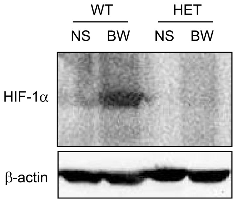
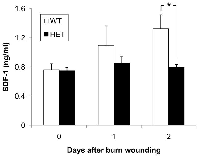
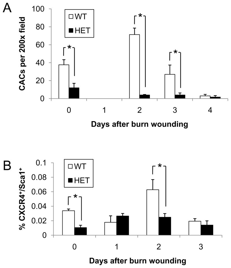
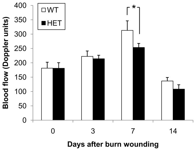
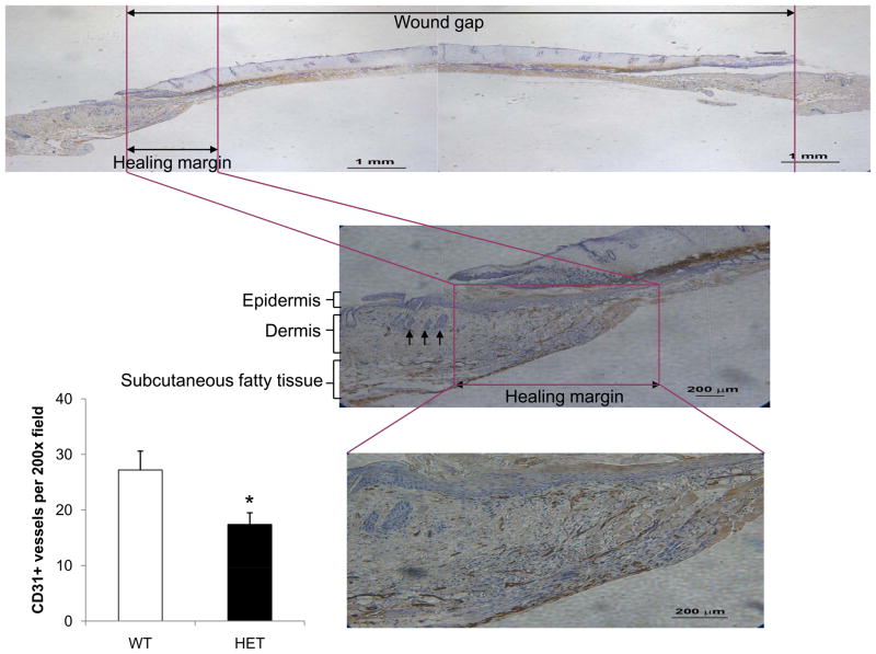
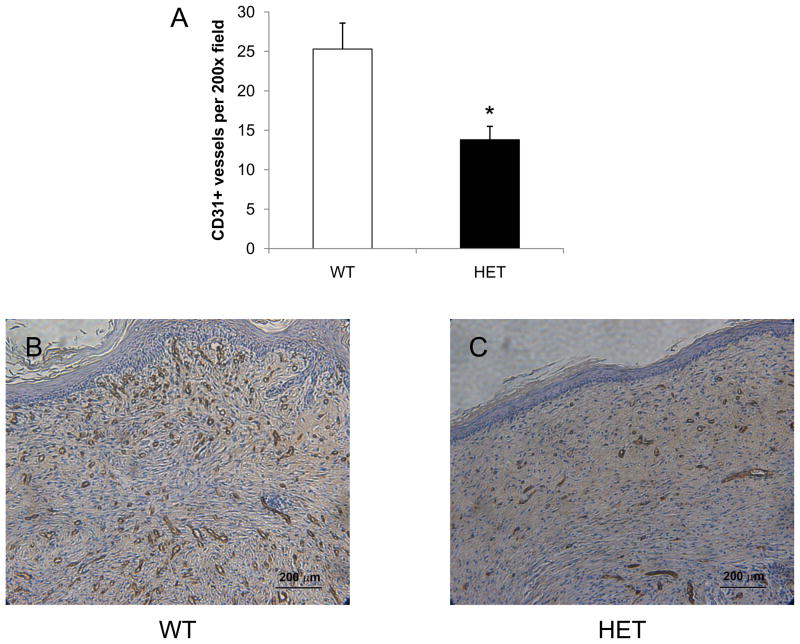
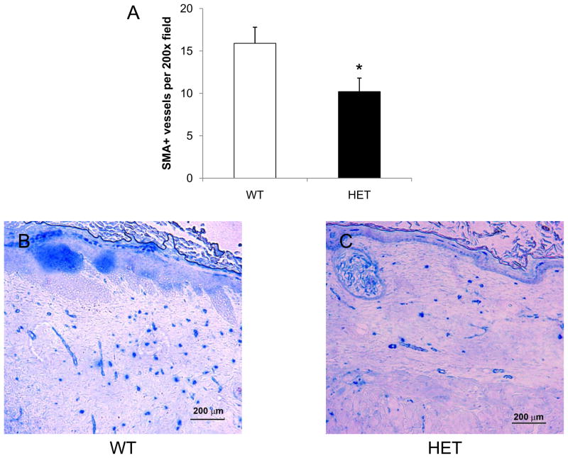
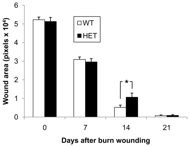
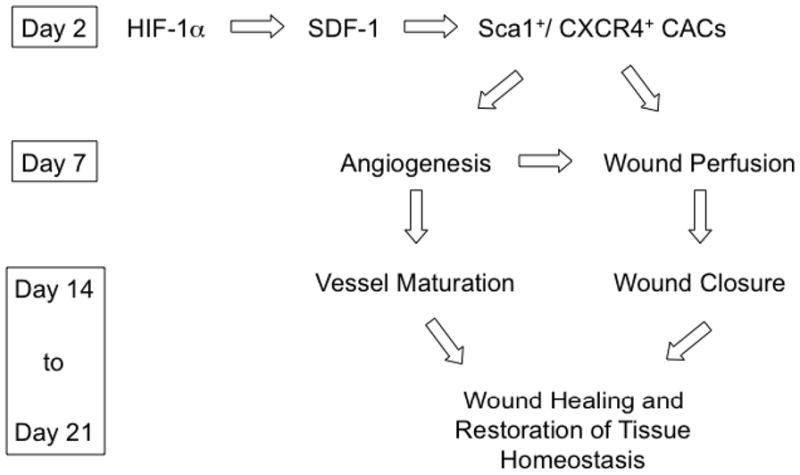
Hypoxia-inducible factor 1 (HIF-1) is a transcription factor that controls vascular responses to hypoxia and ischemia. In this study, mice that were heterozygous (HET) for a null allele at the locus encoding the HIF-1alpha subunit (HET mice) and their wild-type (WT) littermates were subjected to a thermal injury involving 10% of the body surface area. HIF-1alpha protein levels were increased in burn wounds of WT but not of HET mice on day 2. The serum levels of stromal-derived factor 1alpha, which binds to CXCR4, were increased on day 2 in WT but not in HET mice. Circulating angiogenic cells were also increased on day 2 in WT but not in HET mice and included CXCR4(+)Sca1(+) cells. Laser Doppler perfusion imaging demonstrated increased blood flow in burn wounds of WT but not HET mice on day 7. Immunohistochemistry on day 7 revealed a reduced number of CD31(+) vessels at the healing margin of burn wounds in HET as compared with WT mice. Vessel maturation was also impaired in wounds of HET mice as determined by the number of alpha-smooth muscle actin-positive vessels on day 21. The remaining wound area on day 14 was significantly increased in HET mice compared with WT littermates. The percentage of healed wounds on day 14 was significantly decreased in HET mice. These data delineate a signaling pathway by which HIF-1 promotes angiogenesis during burn wound healing.


Conflict of interest statement

Conflict of Interest

The authors state no conflict of interest.

Figures

Figure 1
Figure 1
Analysis of HIF-1α protein levels in burn wounds. Tissue lysates were prepared from the non-burned skin (NS) and burn wound (BW) samples, which were harvested from wild type (WT) and HIF-1α heterozygous-null (HET) mice 2 days after burn wounding. Immunoblot assays were performed using antibodies against HIF-1α and β-actin, which was used as a loading control.
Figure 2
Figure 2
Analysis of serum SDF-1 levels. Peripheral blood samples were collected from WT and HET mice 0, 1 and 2 days after burn wounding. An ELISA for SDF-1 was performed on serum samples (n = 4–9 for each condition). *P<0.05, ANOVA with Tukey Test.
Figure 3
Figure 3
Circulating angiogenic cells in WT and HET mice. (A) Peripheral blood samples were collected from WT and HET mice 0, 1, 2, 3, and 4 days after burn wounding. Blood samples from 3 mice per genotype were pooled for isolation of mononuclear cells, which were cultured in the presence of endothelial growth factors for 4 days, and the number of cells per 200x field which showed uptake of DiI-AcLDL and binding of FITC-BS-1 was determined. The mean number of CACs (± SEM, n = 3 pools for each condition) is shown; *P<0.05, ANOVA with Tukey test. (B) Peripheral blood samples were collected from WT and HET mice 0, 1, 2, and 3 days after burn wounding. The percentage of mononuclear cells that co-expressed CXCR4 and Sca1 was determined by flow cytometry (n = 4 for each condition); *P<0.05, ANOVA with Tukey test.
Figure 4
Figure 4
Burn wound blood flow in WT and HET mice. Laser Doppler perfusion imaging of burn wounds was performed on days 0, 3, 7 and 14 (mean ± SEM, n = 16 for each condition). *P<0.05, ANOVA with Tukey test.
Figure 5
Figure 5
Burn wound vascularization in WT and HET mice on day 7. Immunohistochemical staining for PECAM-1 (CD31) was performed at the healing margin of granulation tissue in each burn wound. In each panel, the healing margin with necrotic burn wound adjacent is outlined in purple. Arrows in the middle panel indicate hair follicles, which identify the normal dermis adjacent to the wound. The bar graph at lower left shows the mean number of CD31+ blood vessels per 200x field (bottom panel) in the healing margin (± SEM, n = 5 for each genotype). *P<0.05, ANOVA with Tukey test.
Figure 6
Figure 6
Analysis of CD31+ vessels in burn wounds on day 21. Immunohistochemical staining for PECAM-1 (CD31) was performed at the healed center of each wound from WT (B) and HET (C) mice and the mean number of stained vessels (± SEM, n = 12 for each genotype) is shown (A). *P<0.05 vs WT, ANOVA with Tukey test.
Figure 7
Figure 7
Analysis of SMA+ vessels in burn wounds on day 21. Immunohistochemical staining for SMA was performed at the healed center of each wound from WT (B) and HET (C) mice and the mean number of stained vessels (± SEM, n = 12 for each genotype) is shown (A). *P<0.05 vs WT, ANOVA with Tukey test.
Figure 8
Figure 8
Kinetics of burn wound healing in WT and HET mice. The bar graph shows the wound area measured by computer-assisted planimetry on days 0, 7, 14 and 21 (mean ± SEM, n = 16 for each condition); *P<0.05 vs WT, ANOVA with Tukey test. Inset shows the percentage of wounds with >95% healing on day 14; *P<0.01 vs WT, χ2 test.
Figure 9
Figure 9
Role of HIF-1 in burn wound vascularization and healing.

References

    1. Brigham PA, McLoughlin E. Burn incidence and medical care use in the United States: estimates, trends, and data sources. J Burn Care Rehabil. 1996;17:95–107. - PubMed
    1. Kerby JD, McGwin G, Jr, George RL, Cross JA, Chaudry IH, Rue LW., 3rd Sex differences in mortality after burn injury: results of analysis of the National Burn Repository of the American Burn Association. J Burn Care Res. 2006;27:452–6. - PubMed
    1. Tredget EE. The basis of fibrosis and wound healing disorders following thermal injury. J Trauma. 2007;62:S69. - PubMed
    1. Folkman J. Is angiogenesis an organizing principle in biology and medicine? J Pediatr Surg. 2007;42:1–11. - PubMed
    1. Semenza GL. Vasculogenesis, angiogenesis, and arteriogenesis: mechanisms of blood vessel formation and remodeling. J Cell Biochem. 2007;102:840–7. - PubMed

Publication types