. 2010 Feb 17:3:3.
doi: 10.1186/1755-1536-3-3.

Insulin-like growth factor binding protein 5 enhances survival of LX2 human hepatic stellate cells

Affiliations

Insulin-like growth factor binding protein 5 enhances survival of LX2 human hepatic stellate cells

Aleksandar Sokolović et al. Fibrogenesis Tissue Repair. .

Abstract

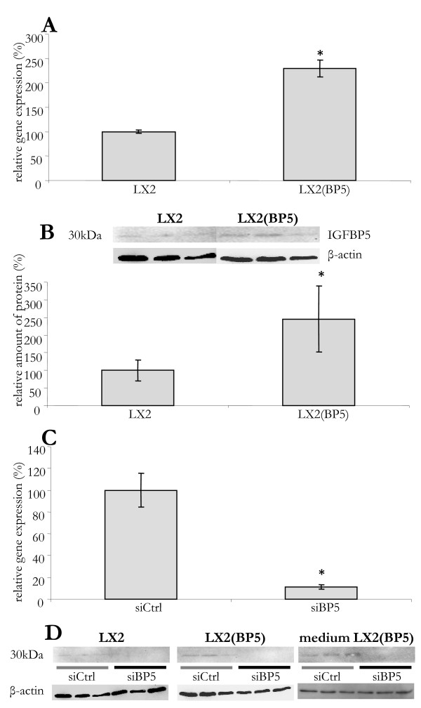
Background: Expression of insulin-like growth factor binding protein 5 (IGFBP5) is strongly induced upon activation of hepatic stellate cells and their transdifferentiation into myofibroblasts in vitro. This was confirmed in vivo in an animal model of liver fibrosis. Since IGFBP5 has been shown to promote fibrosis in other tissues, the aim of this study was to investigate its role in the progression of liver fibrosis.

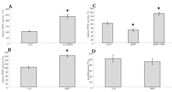
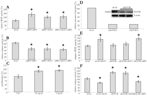
Methods: The effect of IGFBP5 was studied in LX2 cells, a model for partially activated hepatic stellate cells, and in human primary liver myofibroblasts. IGFBP5 signalling was modulated by the addition of recombinant protein, by lentiviral overexpression, and by siRNA mediated silencing. Furthermore, the addition of IGF1 and silencing of the IGF1R was used to investigate the role of the IGF-axis in IGFBP5 mediated effects.

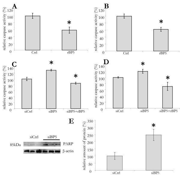
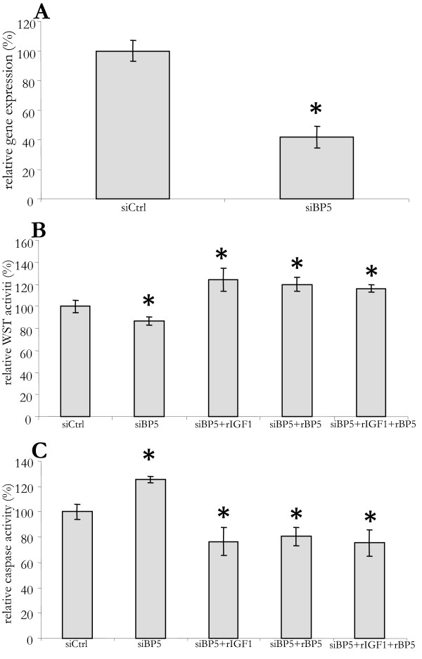
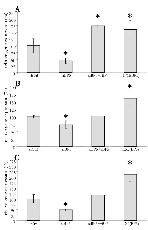
Results: IGFBP5 enhanced the survival of LX2 cells and myofibroblasts via a >50% suppression of apoptosis. This effect of IGFBP5 was not modulated by the addition of IGF1, nor by silencing of the IGF1R. Additionally, IGFBP5 was able to enhance the expression of established pro-fibrotic markers, such as collagen Ialpha1, TIMP1 and MMP1.

Conclusion: IGFBP5 enhances the survival of (partially) activated hepatic stellate cells and myofibroblasts by lowering apoptosis via an IGF1-independent mechanism, and enhances the expression of profibrotic genes. Its lowered expression may, therefore, reduce the progression of liver fibrosis.


Figures

Figure 1
Figure 1
Modulation of IGFBP5 expression in LX2 cells. (A) IGFBP5 mRNA levels in control cells (LX2) and in cells with lentiviral overexpression of IGFBP5 (LX2(BP5)). RNA levels normalized to 36B4 are shown as a percentage of control cells. (B) IGFBP5 protein levels in control (LX2) and transduced (LX2(BP5)) cells as demonstrated on a Western blot (upper panel) and quantified using the LumiImager (lower panel). (C) Silencing of IGFBP5 expression. Cells were transfected with small interfering (si) RNA for IGFBP5 (siBP5) or with a control siRNA (siCtrl). RNA was isolated 48 h later and IGFBP5 mRNA levels were quantified, normalized to 36B4 and given as a percentage of control cells. (D) Proteins were isolated from the cells and the medium 48 h after small interfering RNA (sRNA) transfection. The presence of IGFBP5 protein was visualized by Western blotting in LX2 and LX2(BP5) cells transfected with control siRNA (siCtrl) and IGFBP5 siRNA (siBP5) and in medium obtained from transfected LX2(BP5). The results are given as a percentage of the control cells. The error bars represent standard deviations, with asterisks denoting significant difference (P < 0.05).
Figure 2
Figure 2
IGFBP5 increases survival of LX2 cells. (A) The viability of IGFBP5 overexpressing (LX2(BP5)) and control cells (LX2) cultured in serum-free medium for 48 h was measured using WST. (B) The viability of LX2 cells cultured in serum-free medium for 48 h was determined using WST. At t = 24 and 45 h, recombinant IGFBP5 was added (rBP5). (C) LX2 cells were transfected with small interfering control (siCtrl) or IGFBP5 siRNA (siBP5). At 16 h after transfection the medium was replaced by serum-free medium and 48 h later the viability of the cells was measured using WST. rIGFBP5 was added at t = 24 and 45 h of serum-free culturing (siBP5+rBP5). (D) Bromo-2'-deoxy-uridine (BrdU) incorporation was used to determine the proliferation of LX2 cells cultured in serum-free medium for 48 h. rIGFBP5 was added at t = 24 and 45 h (rBP5). BrdU was added at t = 32 h. The results are shown as percentage of the control cells.
Figure 3
Figure 3
IGFBP5 impact on apoptosis in LX2 cells. (A) LX2 cells were cultured in serum-free medium for 48 h. Recombinant IGFBP5 (rBP5) was added at 24 h and 45 h and, at 48 h, a Caspase 3/7 assay was performed. (B) Cells grown in 10% fetal calf serum were treated with rBP5 as above, with the addition of 0.5 μM gliotoxin after 45 h. (C) LX2 cells were transfected with small interfering control (siCtrl) or IGFBP5 siRNA (siBP5). Sixteen hours after transfection, the medium was replaced by serum-free medium and cells were cultured for additional 48 h before performing the caspase assay. To siIGFBP5 transfected cells, recombinant IGFBP5 was added at t = 24 h and 45 h (siBP5+rBP5). (D) To cells treated with si- and r-BP5 as in C, 0.5 μM gliotoxin was added 3 h before performing the caspase assay. All results are shown as a percentage of the control cells. (E) Apoptosis was induced in LX2 cells transfected with control siRNA (siCtrl) or IGFBP5 siRNA (siBP5) by the addition of 0.5 uM gliotoxin at t = 45 h. Proteins were isolated at 48 h and Western blotting was performed for poly(ASP-ribose) polymerase detection (left panel). Quantification is shown in the right panel. β-actin was used as a loading control.
Figure 4
Figure 4
In human myofibroblasts IGFBP5 silencing decreases survival and promotes apoptosis. (A) Primary human myofibroblasts were transfected with small interfering (si) RNA for IGFBP5 (siBP5) or with a control siRNA (siCtrl). At 16 h after transfection the medium was replaced by serum-free medium. Cells were cultured for an additional 48 h. Then RNA was isolated and IGFBP5 mRNA level determined by quantitative polymerase chain reaction and normalized with 36B4. (B) Recombinant IGFBP5 (rBP5), IGF1 (rIGF1), or both (rIGF1+ rBP5), where added at t = 24 h and 45 h, after replacing the medium, to siBP-5 transfected cells. At 48 h the WST assay was performed. (C) To cells treated as above but grown in 10% fetal calf serum, 0,5 μM gliotoxin was added at 45 h. Caspase 3/7 assay was performed at 48 h. Results are given as a percentage of control cells.
Figure 5
Figure 5
IGFBP5 influences BCL2 expression. LX2 cells were transfected with control small interfering RNA (siCtrl) or IGFBP5 siRNA (siBP5). Sixteen hours after transfection the medium was replaced by serum-free medium. Recombinant IGFBP5 was added at t = 24 h and 45 h, after replacing the medium, to siIGFBP5 transfected cells (siBP5+rBP5). After 48 h RNA was isolated from control (siCtrl), silenced (siBP5), silenced treated with rIGFBP5 (siBP5+rBP5) and LX2 cells overexpression IGFBP5 (LX2(BP5)) and used for quantitative polymerase chain reaction in order to determine BCL2 mRNA. The BCL2 mRNA levels were normalized to 36B4 and given as a percentage of control cells.
Figure 6
Figure 6
Cell survival by IGFBP5 is not affected by IGF1 or mediated by IGF1R. (A) LX2 cells were transfected with IGFBP5 small interfering RNA (siBP5). Sixteen hours after transfection the medium was replaced by serum-free medium for 48 h and then the viability was determined by performing a WST assay. Recombinant IGFBP5 (rBP5), IGF1 (rIGF1), or both (rIGF1+ rBP5), were added at 24 hand 45 h. (B) To cells treated as above, but cultured in 10% fetal calf serum, 0.5 μM gliotoxin was added at 45 h followed by a Caspase 3/7 assay 3 h later. (C) LX2 cells were cultured in serum-free medium for 48 h. rIGF1 was added in different concentrations at 24 h and 45 h, bromo-2'-deoxy-uridine (BrdU) was added at 32 h. Cells were lysed 16 h later (at 48 h) and assayed for BrdU incorporation. (D) LX2 cells were transfected with control (siCtrl) or siRNA for IGF1R (siIGF1R). Sixteen hours after transfection the medium was replaced by serum-free medium and cells were cultured for additional 48 h. The cells were then lysed and used for Western blot analysis in order to detect IGF1R. (E) Cells were transfected and cultured as in D. rIGF1 or rIGFBP5 (rBP5) was added at t = 24 h and 45 h after replacing medium. After 48 h a WST assay was performed. (F) 0.5 μM gliotoxin was added at 45 h, after replacing the medium. Caspase activity was measured 3 h later. All results are given as a percentage control cells.
Figure 7
Figure 7
IGFBP5 influences the expression of genes involved in fibrogenesis. LX2 cells were transfected with small interfering control (siCtrl) or IGFBP5 siRNA (siBP5). Sixteen hours after transfection, medium was replaced by serum-free medium. Recombinant IGFBP5 (rIGFBP5) was added at 24 h and 45 h, after replacing the medium, to siIGFBP5 transfected cells (siBP5+rBP5). After 48 h of culturing on serum-free medium RNA was isolated from control, transfected and transfected treated with rIGFBP5. For comparison, RNA was isolated from LX2 cells overexpressing IGFBP5 cultured in serum-free medium (LX2(BP5)). Collagen 1α1 (A),TIMP1 (B) and MMP1 (C) mRNA levels were measured with quantitative polymerase chain reaction, normalized to 36B4 and given as a percentage of control cells.

Similar articles

Cited by

References

    1. Friedman SL. Liver fibrosis -- from bench to bedside. J Hepatol. 2003;38(Suppl 1):S38–S53. doi: 10.1016/S0168-8278(02)00429-4. - DOI - PubMed
    1. Blomhoff R, Wake K. Perisinusoidal stellate cells of the liver: important roles in retinol metabolism and fibrosis 6. FASEB J. 1991;5:271–277. - PubMed
    1. Wynn TA. Cellular and molecular mechanisms of fibrosis. J Pathol. 2008;214:199–210. doi: 10.1002/path.2277. - DOI - PMC - PubMed
    1. Ikeda K, Wakahara T, Wang YQ, Kadoya H, Kawada N, Kaneda K. In vitro migratory potential of rat quiescent hepatic stellate cells and its augmentation by cell activation. Hepatology. 1999;29:1760–1767. doi: 10.1002/hep.510290640. - DOI - PubMed
    1. Kinnman N, Hultcrantz R, Barbu V, Rey C, Wendum D, Poupon R, Housset C. PDGF-mediated chemoattraction of hepatic stellate cells by bile duct segments in cholestatic liver injury. Lab Invest. 2000;80:697–707. - PubMed