. 2010 Feb 17:10:48.
doi: 10.1186/1471-2148-10-48.

Accelerated microevolution in an outer membrane protein (OMP) of the intracellular bacteria Wolbachia

Affiliations

Accelerated microevolution in an outer membrane protein (OMP) of the intracellular bacteria Wolbachia

Laura Baldo et al. BMC Evol Biol. .

Abstract

Background: Outer membrane proteins (OMPs) of Gram-negative bacteria are key players in the biology of bacterial-host interactions. However, while considerable attention has been given to OMPs of vertebrate pathogens, relatively little is known about the role of these proteins in bacteria that primarily infect invertebrates. One such OMP is found in the intracellular bacteria Wolbachia, which are widespread symbionts of arthropods and filarial nematodes. Recent experimental studies have shown that the Wolbachia surface protein (WSP) can trigger host immune responses and control cell death programming in humans, suggesting a key role of WSP for establishment and persistence of the symbiosis in arthropods.

Results: Here we performed an analysis of 515 unique alleles found in 831 Wolbachia isolates, to investigate WSP structure, microevolution and population genetics. WSP shows an eight-strand transmembrane beta-barrel structure with four extracellular loops containing hypervariable regions (HVRs). A clustering approach based upon patterns of HVR haplotype diversity was used to group similar WSP sequences and to estimate the relative contribution of mutation and recombination during early stages of protein divergence. Results indicate that although point mutations generate most of the new protein haplotypes, recombination is a predominant force triggering diversity since the very first steps of protein evolution, causing at least 50% of the total amino acid variation observed in recently diverged proteins. Analysis of synonymous variants indicates that individual WSP protein types are subject to a very rapid turnover and that HVRs can accommodate a virtually unlimited repertoire of peptides. Overall distribution of WSP across hosts supports a non-random association of WSP with the host genus, although extensive horizontal transfer has occurred also in recent times.

Conclusions: In OMPs of vertebrate pathogens, large recombination impact, positive selection, reduced structural and compositional constraints, and extensive lateral gene transfer are considered hallmarks of evolution in response to the adaptive immune system. However, Wolbachia do not infect vertebrates. Here we predict that the rapid turnover of WSP loop motifs could aid in evading or inhibiting the invertebrate innate immune response. Overall, these features identify WSP as a strong candidate for future studies of host-Wolbachia interactions that affect establishment and persistence of this widespread endosymbiosis.


Figures

Figure 1
Figure 1
WSP gene structure. (A) Schematic representation of WSP structure depicting the signal peptide (SP), the four hypervariable regions (HVRs), interspaced by four conserved regions (CRs). (B) Representatives of most diverse WSP sequences named by a corresponding allele. Sections used for typing include HVR motifs plus short stretches of the two flanking CRs (bracketed below the two alignments).
Figure 2
Figure 2
Predicted folding and localization of WSP in the outer membrane. WSP shows an eight antiparallel stranded β-barrel structure with four highly hydrophilic loops (L1-4) protruding into the extracellular side (OUT). C- and N-termini are in the periplasmic side (IN). Loop size and amino acid content greatly vary among WSP sequences, thus exact folding and localization for these extracellular regions cannot be reliably predicted.
Figure 3
Figure 3
Clusters of closely related WSP proteins (n = 252) estimated by eBURST. Circles correspond to distinct proteins; area of the circles is proportional to the number of allele variants coding for a protein. Clusters of linked proteins (i.e. WSP complexes C1-57) are identified as groups of proteins sharing three out of four HVR haplotypes (SHVs) with a primary founder (in blue); subgroup founders (yellow) connect to double HVR variants (DHV). In red are recombinant proteins within a complex, labeled with the corresponding allele number or a representative allele in case of multiple variants (*). For clarity only complexes with at least three proteins or recombinants were labeled (C1-19, C47, C51). 183 singletons were removed. Refer to Additional file 2 for profile and allele identification within complexes.
Figure 4
Figure 4
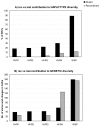
Relative contribution of recombination (rec) versus mutation (mut) to WSP haplotype (A) and genetic (B) diversity. A) Percentage of recombinant and mutant SHVs per class of HVRs and per protein; B) total number of amino acid changes introduced in SHVs by recombination and mutation per class of HVR and per protein. Overall ratio of rec/mut is about 1/8 to WSP haplotype diversity and about 1/1 to WSP amino acid diversity.
Figure 5
Figure 5
Rarefaction curve of the cumulative number of distinct wsp alleles and proteins. Analysis is based on a random resampling of 20. The best curve fit was y = -1.347224537·10-1 x + 1179.975114 x/(x + 1034.575827), with a very small residual of rss = 2.251531074·10-1. There was no good fit to a curve with asymptotic features.
Figure 6
Figure 6
Most widespread wsp alleles and relative host taxonomic distribution. Note that some of the most common alleles are predicted founders of complexes (e.g. wsp-10, 23 and 18). wsp-10 is the most widespread in terms of diversity of host species, genera and families and corresponds to the ancestral allele of C6 (see also Fig. 3 and Table 3).

Similar articles

Cited by

References

    1. Lin J, Huang S, Zhang Q. Outer membrane proteins: key players for bacterial adaptation in host niches. Microbes Infect. 2002;4(3):325–331. doi: 10.1016/S1286-4579(02)01545-9. - DOI - PubMed
    1. Koebnik R, Locher KP, Van Gelder P. Structure and function of bacterial outer membrane proteins: barrels in a nutshell. Mol Microbiol. 2000;37(2):239–253. doi: 10.1046/j.1365-2958.2000.01983.x. - DOI - PubMed
    1. Ohashi N, Zhi N, Zhang Y, Rikihisa Y. Immunodominant major outer membrane proteins of Ehrlichia chaffeensis are encoded by a polymorphic multigene family. Infect Immun. 1998;66(1):132–139. - PMC - PubMed
    1. Mes TH, van Putten JP. Positively selected codons in immune-exposed loops of the vaccine candidate OMP-P1 of Haemophilus influenzae. J Mol Evol. 2007;64(4):411–422. doi: 10.1007/s00239-006-0021-2. - DOI - PMC - PubMed
    1. Vandeputte-Rutten L, Bos MP, Tommassen J, Gros P. Crystal structure of Neisserial surface protein A (NspA), a conserved outer membrane protein with vaccine potential. J Biol Chem. 2003;278(27):24825–24830. doi: 10.1074/jbc.M302803200. - DOI - PubMed

Publication types

MeSH terms

Substances

LinkOut - more resources