

Use of ultra-high-resolution optical coherence tomography to detect in vivo characteristics of Descemet's membrane in Fuchs' dystrophy
- PMID: 20163865
- PMCID: PMC2881170
- DOI: 10.1016/j.ophtha.2009.10.027
Use of ultra-high-resolution optical coherence tomography to detect in vivo characteristics of Descemet's membrane in Fuchs' dystrophy
Abstract
Purpose: To demonstrate the capability of ultra-high-resolution (UHR) anterior segment optical coherence tomography (OCT) to image Descemet's membrane (DM) and measure its thickness in vivo. (2) To evaluate the use of DM characteristics and thickness in the diagnosis of Fuchs' dystrophy.
Design: Case-control study.
Participants: Twenty eyes of 12 Fuchs' dystrophy patients, 20 eyes of 13 young normal, and 20 eyes of 15 elderly normal subjects.
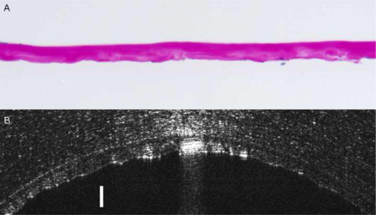
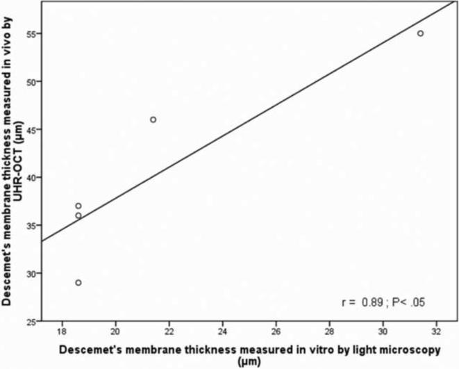
Methods: Subjects were imaged using novel, custom-built UHR-OCT. Images were used to describe the characteristics of DM. Custom-made software was used to measure DM thickness and central corneal thickness (CCT). Specimens of DM obtained from Fuchs' dystrophy patients who underwent endothelial keratoplasty (EK) were histopathologically examined. Regression analyses were used to assess the correlation of DM thickness measured by UHR-OCT in vivo and by light microscopy and to determine the intergroup correlations between age, CCT, and DM thickness.
Main outcome measures: We assessed DM characteristics and thickness, CCT, and age.
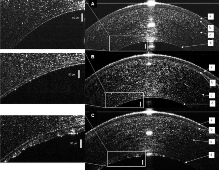
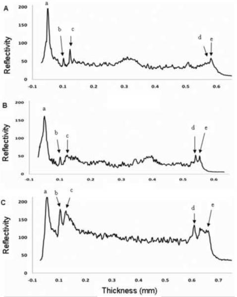
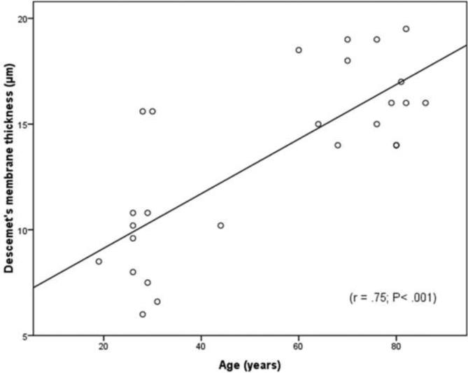
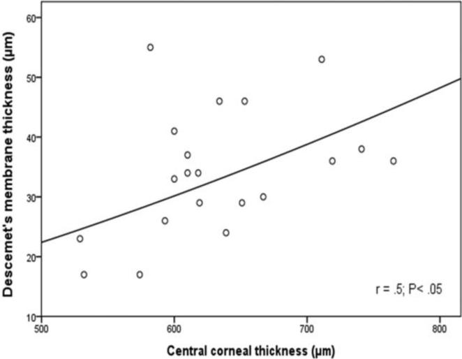
Results: Using UHR-OCT, the DM seemed in normal young subjects as a single, opaque, smooth line and in normal elderly subjects as a band of 2 smooth opaque lines with a translucent space in between. In Fuchs' dystrophy, DM appeared as a thickened band of 2 opaque lines; the anterior line was smooth whereas the posterior line had a wavy and irregular appearance with areas of localized thickenings. The DM thickness measured in vivo by UHR-OCT correlated significantly with that measured by light microscopy in 5 Fuchs' dystrophy eyes that underwent EK. The average central thicknesses of DM in normal young, in normal elderly and in Fuchs' dystrophy eyes were 10+/-3, 16+/-2, and 34+/-11 microm, respectively (P<0.001). There was a significant correlation between age and DM thickness only in normal groups. In Fuchs' dystrophy patients, there was a significant correlation between CCT and DM thickness that was not significant for normal groups.
Conclusions: Ultra-high-resolution OCT is an innovative technique for the in vivo imaging of DM. Determining DM characteristics and thickness by UHR-OCT could be a new approach for the diagnosis of Fuchs' dystrophy.
Copyright 2010 American Academy of Ophthalmology. Published by Elsevier Inc. All rights reserved.
Figures
References
-
- Seitzman GD, Gottsch JD, Stark WJ. Cataract surgery in patients with Fuchs' corneal dystrophy: expanding recommendations for cataract surgery without simultaneous keratoplasty. Ophthalmology. 2005;112:441–6. - PubMed
-
- Seitzman GD. Cataract surgery in Fuchs' dystrophy. Curr Opin Ophthalmol. 2005;16:241–5. - PubMed
-
- Adamis AP, Filatov V, Tripathi BJ, Tripathi RC. Fuchs' endothelial dystrophy of the cornea. Surv Ophthalmol. 1993;38:149–68. - PubMed
-
- Rodrigues MM, Krachmer JH, Hackett J, et al. Fuchs' corneal dystrophy: a clinicopathologic study of the variation in corneal edema. Ophthalmology. 1986;93:789–96. - PubMed
Publication types
MeSH terms
Grants and funding
LinkOut - more resources
Full Text Sources
Research Materials
