

The proteome of Shigella flexneri 2a 2457T grown at 30 and 37 degrees C
- PMID: 20164057
- PMCID: PMC2877981
- DOI: 10.1074/mcp.M900446-MCP200
The proteome of Shigella flexneri 2a 2457T grown at 30 and 37 degrees C
Abstract
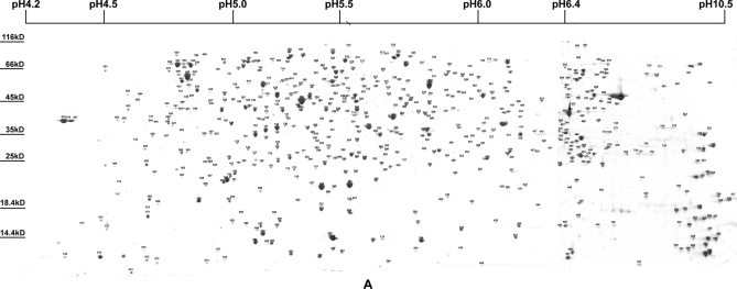
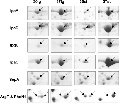
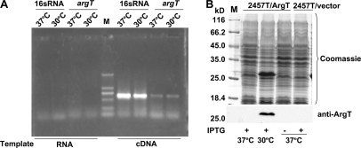
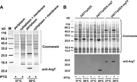
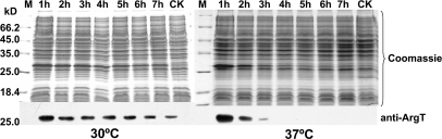
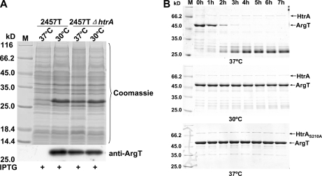
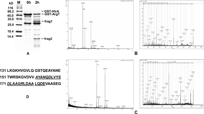
To upgrade the proteome reference map of Shigella flexneri 2a 2457T, the protein expression profiles of log phase and stationary phase cells grown at 30 and 37 degrees C were thoroughly analyzed using multiple overlapping narrow pH range (between pH 4.0 and 11.0) two-dimensional gel electrophoresis. A total of 723 spots representing 574 protein entries were identified by MALDI-TOF/TOF MS, including the majority of known key virulence factors. 64 hypothetical proteins and six misannotated proteins were also experimentally identified. A comparison between the four proteome maps showed that most of the virulence-related proteins were up-regulated at 37 degrees C, and the differences were more notable in stationary phase cells, suggesting that the expressions of these virulence factors were not only controlled by temperature but also controlled by the nutrients available in the environment. The expression patterns of some virulence-related genes under the four different conditions suggested that they might also be regulated at the post-transcriptional level. A further significant finding was that the expression of the protein ArgT was dramatically up-regulated at 30 degrees C. The results of semiquantitative RT-PCR analysis showed that expression of argT was not regulated at the transcriptional level. Therefore, we carried out a series of experiments to uncover the mechanism regulating ArgT levels and found that the differential expression of ArgT was due to its degradation by a periplasmic protease, HtrA, whose activity, but not its synthesis, was affected by temperature. The cleavage site in ArgT was between position 160 (Val) and position 161 (Ala). These results may provide useful insights for understanding the physiology and pathogenesis of S. flexneri.
Figures
Similar articles
-
Analysis of Soluble protein complexes in Shigella flexneri reveals the influence of temperature on the amount of lipopolysaccharide.Mol Cell Proteomics. 2013 May;12(5):1250-8. doi: 10.1074/mcp.M112.025270. Epub 2013 Feb 2. Mol Cell Proteomics. 2013. PMID: 23378524 Free PMC article.
-
Dynamic proteome changes of Shigella flexneri 2a during transition from exponential growth to stationary phase.Genomics Proteomics Bioinformatics. 2007 May;5(2):111-20. doi: 10.1016/S1672-0229(07)60021-7. Genomics Proteomics Bioinformatics. 2007. PMID: 17893076 Free PMC article.
-
[Comparative proteomics research on removing of large invasive plasmid pINV of Shigella flexneri 2a strain 2457T].Wei Sheng Wu Xue Bao. 2005 Jun;45(3):410-4. Wei Sheng Wu Xue Bao. 2005. PMID: 15989237 Chinese.
-
Immunoproteome analysis of soluble and membrane proteins of Shigella flexneri 2457T.World J Gastroenterol. 2006 Nov 7;12(41):6683-8. doi: 10.3748/wjg.v12.i41.6683. World J Gastroenterol. 2006. PMID: 17075984 Free PMC article.
-
Shigella flexneri: genetics of entry and intercellular dissemination in epithelial cells.Curr Top Microbiol Immunol. 1994;192:217-41. doi: 10.1007/978-3-642-78624-2_10. Curr Top Microbiol Immunol. 1994. PMID: 7859507 Review. No abstract available.
Cited by
-
Identification of differential proteomics in Epstein-Barr virus-associated gastric cancer and related functional analysis.Cancer Cell Int. 2021 Jul 12;21(1):368. doi: 10.1186/s12935-021-02077-6. Cancer Cell Int. 2021. PMID: 34247602 Free PMC article.
-
Proteomic analysis of synovial fluid: insight into the pathogenesis of knee osteoarthritis.Int Orthop. 2013 Jun;37(6):1045-53. doi: 10.1007/s00264-012-1768-2. Epub 2013 Mar 28. Int Orthop. 2013. PMID: 23532587 Free PMC article.
-
Combining blue native polyacrylamide gel electrophoresis with liquid chromatography tandem mass spectrometry as an effective strategy for analyzing potential membrane protein complexes of Mycobacterium bovis bacillus Calmette-Guérin.BMC Genomics. 2011 Jan 18;12:40. doi: 10.1186/1471-2164-12-40. BMC Genomics. 2011. PMID: 21241518 Free PMC article.
-
Analysis of the proteome of intracellular Shigella flexneri reveals pathways important for intracellular growth.Infect Immun. 2013 Dec;81(12):4635-48. doi: 10.1128/IAI.00975-13. Epub 2013 Oct 7. Infect Immun. 2013. PMID: 24101689 Free PMC article.
-
Analysis of Soluble protein complexes in Shigella flexneri reveals the influence of temperature on the amount of lipopolysaccharide.Mol Cell Proteomics. 2013 May;12(5):1250-8. doi: 10.1074/mcp.M112.025270. Epub 2013 Feb 2. Mol Cell Proteomics. 2013. PMID: 23378524 Free PMC article.
References
-
- Wei J., Goldberg M. B., Burland V., Venkatesan M. M., Deng W., Fournier G., Mayhew G. F., Plunkett G., 3rd, Rose D. J., Darling A., Mau B., Perna N. T., Payne S. M., Runyen-Janecky L. J., Zhou S., Schwartz D. C., Blattner F. R. (2003) Complete genome sequence and comparative genomics of Shigella flexneri serotype 2a strain 2457T. Infect. Immun 71, 2775–2786 - PMC - PubMed
-
- Liao X., Ying T., Wang H., Wang J., Shi Z., Feng E., Wei K., Wang Y., Zhang X., Huang L., Su G., Huang P. (2003) A two-dimensional proteome map of Shigella flexneri. Electrophoresis 24, 2864–2882 - PubMed
-
- Buchrieser C., Glaser P., Rusniok C., Nedjari H., D'Hauteville H., Kunst F., Sansonetti P., Parsot C. (2000) The virulence plasmid pWR100 and the repertoire of proteins secreted by the type III secretion apparatus of Shigella flexneri. Mol. Microbiol 38, 760–771 - PubMed
-
- Le Gall T., Mavris M., Martino M. C., Bernardini M. L., Denamur E., Parsot C. (2005) Analysis of virulence plasmid gene expression defines three classes of effectors in the type III secretion system of Shigella flexneri. Microbiology 151, 951–962 - PubMed
Publication types
MeSH terms
Substances
LinkOut - more resources
Full Text Sources
