. 2010 Jun;38(10):e115.
doi: 10.1093/nar/gkq042. Epub 2010 Feb 17.

Minimizing off-target signals in RNA fluorescent in situ hybridization

Affiliations

Minimizing off-target signals in RNA fluorescent in situ hybridization

Aaron Arvey et al. Nucleic Acids Res. 2010 Jun.

Abstract

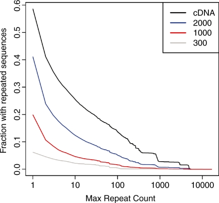
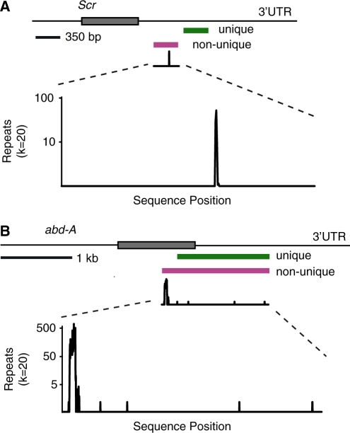
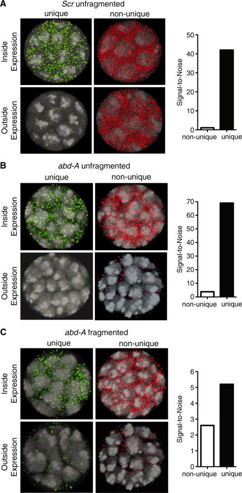
Fluorescent in situ hybridization (FISH) techniques are becoming extremely sensitive, to the point where individual RNA or DNA molecules can be detected with small probes. At this level of sensitivity, the elimination of 'off-target' hybridization is of crucial importance, but typical probes used for RNA and DNA FISH contain sequences repeated elsewhere in the genome. We find that very short (e.g. 20 nt) perfect repeated sequences within much longer probes (e.g. 350-1500 nt) can produce significant off-target signals. The extent of noise is surprising given the long length of the probes and the short length of non-specific regions. When we removed the small regions of repeated sequence from either short or long probes, we find that the signal-to-noise ratio is increased by orders of magnitude, putting us in a regime where fluorescent signals can be considered to be a quantitative measure of target transcript numbers. As the majority of genes in complex organisms contain repeated k-mers, we provide genome-wide annotations of k-mer-uniqueness at http://cbio.mskcc.org/ approximately aarvey/repeatmap.


Figures

Figure 1.
Figure 1.
A large fraction of RNA FISH probes contain short non-unique regions. The Y-axis shows the fraction of D. melanogaster RNA-coding sequences that contain genomic repeated sequences of size 20 nt (20-mers) or greater. Sequences have been grouped into full-length mRNA-coding (cDNA) sequences, or subsets of such coding sequences that are of lengths 2000, 1000 or 300 nt, respectively. The X-axis shows the number of 20-mer repeats that are present at a given probe sequence length. For example, about 15% of full-length cDNA sequences have 20-mer or larger repeats that are found more than 100 times in the genome.
Figure 2.
Figure 2.
Scr and abd-A probes with and without 20-mer repeats. The lines at the top represent mature mRNA sequences for the Scr (A) and abd-A (B) genes, with the positions of probe regions denoted beneath. The shaded boxes represent the open reading frames of the respective mRNAs. Below the non-unique probes is shown the graphical output from the RepeatMap program, set to detect repeated sequences of 20 nt. The Y-axis log10 scale shows the number of times a k-mer repeat (k = 20) is found elsewhere in the D. melanogaster genome. The X-axis plot shows the nucleotide position of the repeat within the probe sequence.
Figure 3.
Figure 3.
Unique probes are qualitatively more specific than non-unique probes. Low-resolution images are shown for embryos hybridized with (A) Scr unique probe, (B) Scr non-unique probe, (C) abd-A unfragmented unique probe (D) abd-A unfragmented non-unique probe (E) abd-A fragmented unique probe (F) abd-A fragmented non-unique probe. The locations of the probes are shown in Figure 2. Embryos are shown anterior to the left, dorsal up. White circles mark the locations of the areas that were imaged at higher resolution and that are shown in Figure 4.
Figure 4.
Figure 4.
Unique probes are quantitatively much more specific than non-unique probes. High-resolution images are shown for regions inside and outside canonical regions of expression for Scr and abd-A (areas of white circles in Figure 3) for both unique and non-unique probes. Images show stains for (A) Scr, (B) abd-A (unfragmented) and (C) abd-A (fragmented). The DAPI (nuclear stain) is shown in light gray. Although unique and non-unique probes are shown with green and red signals, respectively, both probes were labeled with the same hapten, DIG, for quantitative measurements. The panel adjacent to each set of images for the respective probes shows the mean signal-to-noise ratios for each of the probes and indicates that non-unique probes are dramatically less specific. The quantitative data represents results from ∼10 embryos from three different hybridization experiments.

References

    1. Kosman D, Mizutani C, Lemons D, Cox W, McGinnis W, Bier E. Multiplex detection of RNA expression in Drosophila embryos. Science. 2004;305:846. - PubMed
    1. Gregorieff A, Pinto D, Begthel H, Destrée O, Kielman M, Clevers H. Expression pattern of Wnt signaling components in the adult intestine. Gastroenterology. 2005;129:626–638. - PubMed
    1. Pezo R, Gandhi S, Shirley L, Pestell R, Augenlicht L, Singer R. Single-cell transcription site activation predicts chemotherapy response in human colorectal tumors. Cancer Res. 2008;68:4977–4982. - PMC - PubMed
    1. Levsky J, Singer R. Fluorescence in situ hybridization: past, present and future. J. Cell Sci. 2003;116:2833–2838. - PubMed
    1. Paré A, Lemons D, Kosman D, Beaver W, Freund Y, McGinnis W. Counting Hox transcripts within single cells in fixed Drosophila embryos: evidence for transcriptional bursting. Curr. Biol. 2009;19:1–6. - PMC - PubMed

Publication types

MeSH terms