. 2010 May;298(5):F1130-9.
doi: 10.1152/ajprenal.00618.2009. Epub 2010 Feb 17.

Expression and response of acid-sensing ion channels in urinary bladder to cyclophosphamide-induced cystitis

Affiliations

Expression and response of acid-sensing ion channels in urinary bladder to cyclophosphamide-induced cystitis

Kimberly Corrow et al. Am J Physiol Renal Physiol. 2010 May.

Abstract

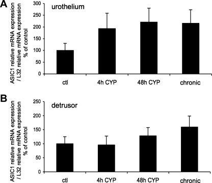
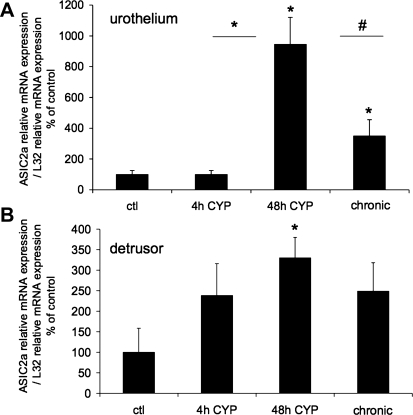
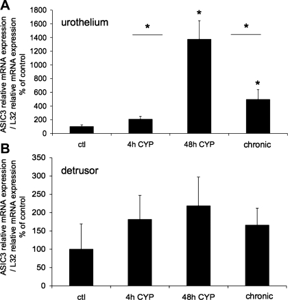
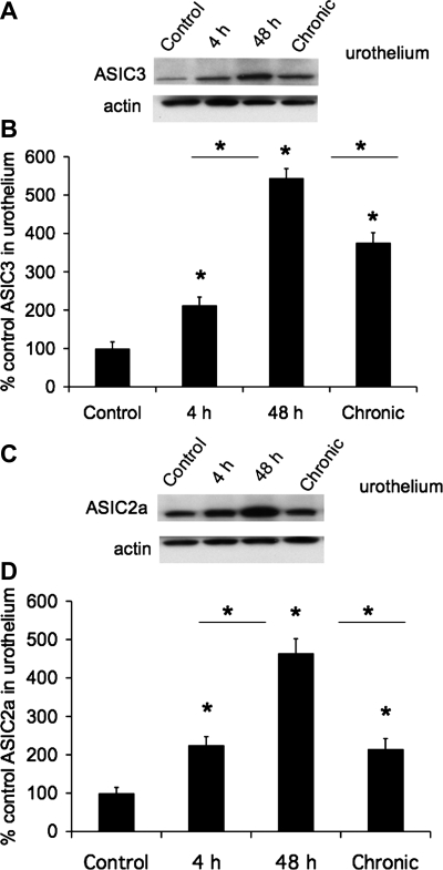
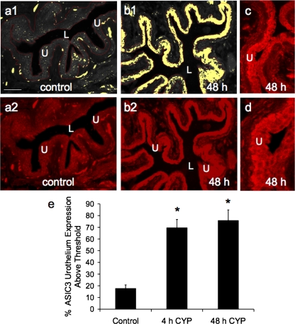
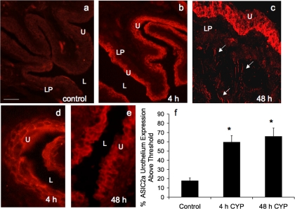
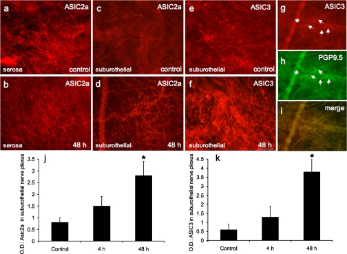
The expression of acid-sensing ion channel (ASIC) isoforms, ASIC1, ASIC2a, and ASIC3, was examined in the urinary bladder after cyclophosphamide (CYP)-induced cystitis of varying duration (4 h, 48 h, and chronic). Immunohistochemical, Western blot, and quantitative PCR approaches were used to evaluate channel expression and effects of CYP-induced cystitis in whole urinary bladder and split-bladder preparations from control (no inflammation) and CYP-treated rats. Quantitative PCR demonstrated significant (P ≤ 0.01) increases in ASIC2a and ASIC3 transcripts with CYP-induced cystitis (48 h and chronic) in the urothelium but no changes (e.g., ASIC3) or modest changes (e.g., ASIC2a) in detrusor smooth muscle. ASIC1 mRNA expression in the urothelium or detrusor was not affected by CYP-induced cystitis. Immunohistochemistry for ASIC2a and ASIC3 protein expression revealed significant (P ≤ 0.01) increases in ASIC immunoreactivity in the urothelium and suburothelial plexus with CYP-induced cystitis at all time points examined. Western blotting for ASIC2a and ASIC3 protein expression was complementary and revealed significant (P ≤ 0.01) increases in ASIC immunoreactivity. For the first time, these studies demonstrate that CYP-induced cystitis alters ASIC2a and ASIC3 expression in the urinary bladder; ASIC1 transcript expression is not altered by CYP-induced cystitis. Future studies are necessary to determine ASIC isoform contributions to micturition reflexes in control and inflamed urinary bladder.


Figures

Fig. 1.
Fig. 1.
Acid-sensing ion channel (ASIC) isoform 1 (ASIC1) mRNA expression in urothelium and detrusor in control rats and after 4 h (h), 48 h, and chronic (10 days) cyclophosphamide (CYP)-induced bladder inflammation (n = 5–7 for each group). A and B: summary histograms of relative expression of the ASIC1 transcript in urothelium and detrusor as a percentage of control and normalized to relative expression of the housekeeping gene L32 obtained from quantitative PCR. No differences in ASIC1 expression with CYP-induced cystitis of any duration compared with control in urothelium + suburothelium (A) or detrusor smooth muscle (B) were detected (P = 0.64–0.76).
Fig. 2.
Fig. 2.
ASIC2a mRNA expression in urothelium and detrusor in control rats and after 4 h, 48 h, and chronic (10 days) CYP-induced bladder inflammation (n = 5–7 for each group). A and B: summary histograms of relative expression of the ASIC2a transcript in urothelium and detrusor as a percentage of control and normalized to relative expression of the housekeeping gene L32 obtained from quantitative PCR. *P ≤ 0.01; #P ≤ 0.05 vs. control or as indicated between groups by horizontal lines.
Fig. 3.
Fig. 3.
ASIC3 mRNA expression in urothelium and detrusor in control rats and after 4 h, 48 h, and chronic (10 days) CYP-induced bladder inflammation (n = 5–7 for each group). A and B: summary histograms of relative expression of the ASIC3 transcript in urothelium and detrusor as a percentage of control and normalized to relative expression of the housekeeping gene L32 obtained from quantitative PCR. *P ≤ 0.01 vs. control or as indicated between groups by horizontal lines.
Fig. 4.
Fig. 4.
Increased ASIC3 and ASIC2a protein expression in urothelium with CYP-induced cystitis (4 h, 48 h, and chronic). A: Western blot of urothelium (20 μg) for ASIC3 expression in control rats and those treated with CYP for 4 h, 48 h, and 10 days (chronic). Actin expression was used as a loading control. B: relative urothelium ASIC3 band density in all groups examined normalized to actin in the same samples presented as a percentage of control ASIC3 expression. C: Western blot of urothelium (20 μg) for ASIC2a expression in control rats and those treated with CYP for 4 h, 48 h, and 10 days (chronic). Actin expression was used as a loading control. D: relative urothelium ASIC2a band density in all groups examined normalized to actin in the same samples presented as a percentage of control ASIC2a expression. *P ≤ 0.01 vs. control or as indicated between groups by horizontal lines (n = 5–6 for each group).
Fig. 5.
Fig. 5.
Semiquantitative analysis of ASIC3 immunoreactivity (IR) in the urothelium after CYP-induced cystitis. a1 and b1: gray-scale versions of ASIC3-IR in control urinary bladder and 48 h after CYP treatment, with urothelium (U) outlined in red. A threshold encompassing an intensity range of 100–250 gray-scale values was applied to the region of interest (a1 and b1); the same threshold was subsequently used for all images. Percent ASIC expression above threshold in the total area selected was then determined. Little ASIC3-IR (absence of yellow within the outlined region) is above threshold in control bladders (a1) compared with significant ASIC3-IR after 48 h of CYP treatment (b1, presence of yellow within U). a2 and b2: fluorescence images corresponding to gray-scale images (a1 and b1). c and d: higher-power images of ASIC3-IR in the urothelium after 48 h of CYP treatment. Calibration bar represents 50 μm. L, lumen. e: ASIC3 expression in the urothelium, with CYP treatment expressed as a percentage of control averaged for all bladders from all conditions examined (n = 6). *P ≤ 0.01.
Fig. 6.
Fig. 6.
ASIC2a-IR in cryostat sections of CYP-treated urothelium. CYP treatment [4 h (b and d) and 48 h (c and e)] significantly (P ≤ 0.05) increased the percentage of ASIC2a-IR in urothelium compared with control (a). c: confocal z-stack image demonstrating ASIC2a-IR in urothelium and diffuse ASIC2a in lamina propria (LP) and in putative nerve fibers (arrows). d and e: higher-power fluorescence images of ASIC2a-IR in urothelium after 4 h and 48 h of CYP-induced cystitis. For all images, exposure times were held constant, and all tissues were processed simultaneously. In rats treated with CYP for 4 and 48 h, ASIC2a expression was visible in urothelium (b–e), whereas control (a) urinary bladder showed little or no ASIC2a-IR. Calibration bar represents 50 μm in a–c and 25 μm in d and e. L, lumen. f: ASIC2a expression above threshold in urothelium of CYP-treated (4 h and 48 h) rats expressed as a percentage of control. Semiquantitative analyses were performed as described in Fig. 5 legend. CYP treatment (4 h and 48 h) significantly (P ≤ 0.01) increased ASIC2a-IR in urothelium (n = 6 for each group).
Fig. 7.
Fig. 7.
ASIC2a and ASIC3 expression in nerve fibers in urinary bladder. Fluorescence photographs show ASIC2a- or ASIC3-IR nerve fibers of suburothelial nerve plexus in whole-mount preparations of urothelium in control (c and e) and CYP-treated (48 h) rats (d and f). ASIC2a expression was also present in presumptive nerve fibers on the serosal surface of urinary bladder (a), but expression was not regulated by CYP treatment (b). ASIC3-IR nerve fibers in suburothelial nerve plexus expressed the pan-neuronal marker protein gene product (PGP) 9.5. g: ASIC3 expression in nerve fibers (arrows) of suburothelial nerve plexus in CYP-treated (48 h) rats. h: whole-mount preparation in a stained for PGP9.5, with ASIC3-IR nerve fibers expressing PGP9.5 (arrows). i: merged image demonstrating overlap between ASIC and PGP immunostaining in suburothelial nerve plexus. j and k: optical density (OD) of ASIC2a and ASIC3 expression in suburothelial nerve plexus. CYP treatment (48 h) significantly (P ≤ 0.01) increased ASIC2a (d and j) and ASIC3 (f and k) expression in suburothelial nerve plexus. ∗ in g and h shows a larger-caliber nerve fiber out of the focal plane. Calibration bar represents 80 μm in a–i.

Similar articles

Cited by

References

    1. Abramoff MD, Magelhaes PJ, Ram SJ. Image processing with ImageJ. Biophotonics Int 11: 36–42, 2004
    1. Arms L, Girard BM, Vizzard MA. Expression and function of CXCL12/CXCR4 in rat urinary bladder with cyclophosphamide-induced cystitis. Am J Physiol Renal Physiol 298: F589–F600, 2010 - PMC - PubMed
    1. Birder LA. More than just a barrier: urothelium as a drug target for urinary bladder pain. Am J Physiol Renal Physiol 289: F489–F495, 2005 - PubMed
    1. Birder LA. Urinary bladder urothelium: molecular sensors of chemical/thermal/mechanical stimuli. Vascul Pharmacol 45: 221–226, 2006 - PubMed
    1. Braas KM, May V, Zvara P, Nausch B, Kliment J, Dunleavy JD, Nelson MT. Role for pituitary adenylate cyclase activating polypeptide (PACAP) in cystitis-induced plasticity of micturition reflexes. Am J Physiol Regul Integr Comp Physiol 290: R951–R962, 2006 - PMC - PubMed

Publication types

LinkOut - more resources