. 2010 Apr 16;285(16):12232-40.
doi: 10.1074/jbc.M109.065920. Epub 2010 Feb 17.

A novel LZAP-binding protein, NLBP, inhibits cell invasion

Affiliations

A novel LZAP-binding protein, NLBP, inhibits cell invasion

Junhye Kwon et al. J Biol Chem. .

Abstract

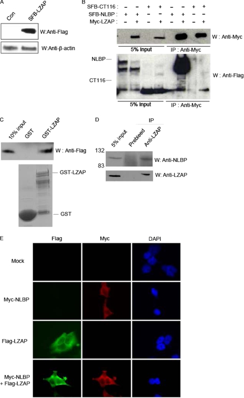
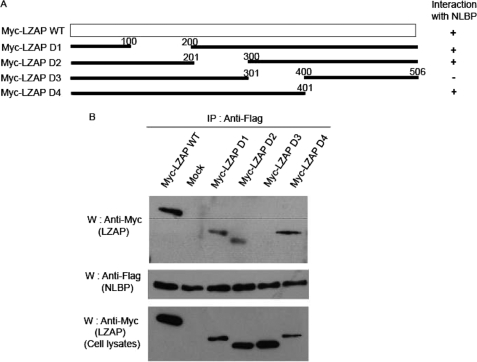
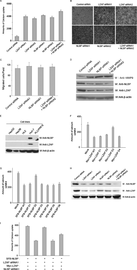
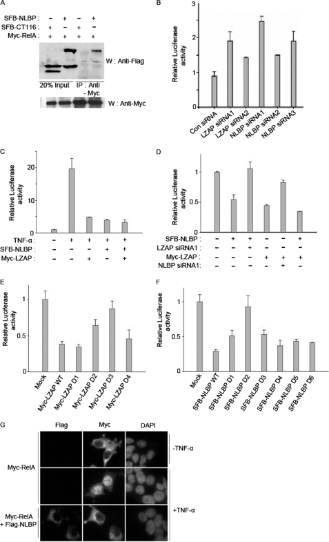
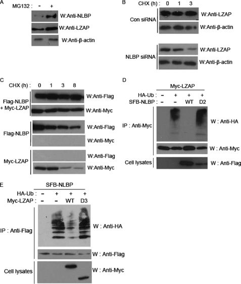
LXXLL/leucine zipper-containing alternative reading frame (ARF)-binding protein (LZAP) was recently shown to function as a tumor suppressor through inhibition of the NF-kappaB signaling pathway. LZAP is also known as a negative regulator of cell invasion, and its expression was demonstrated to be reduced in several tumor tissues. However, the molecular mechanism of the negative effect of LZAP on cell invasion is unclear. In this study, we identify NLBP as a novel LZAP-binding protein using tandem affinity purification. We demonstrate the negative effects of NLBP on cell invasion and the NF-kappaB signaling pathway. NLBP expression was not detected in hepatocellular carcinoma cells with strong invasive activity, whereas its expression was detected in a hepatocellular carcinoma cell line with no invasive activity. We also demonstrate that these two proteins mutually affect the stability of each other by inhibiting ubiquitination of the other protein. Based on these results, we suggest that NLBP may act as a novel tumor suppressor by inhibiting cell invasion, blocking NF-kappaB signaling, and increasing stability of the LZAP protein.


Figures

FIGURE 1.
FIGURE 1.
Identification of NLBP as a novel LZAP-binding protein. A, establishment of human embryonic kidney 293T cell lines stably expressing SFB-LZAP. Cell extracts prepared from 293T cells stably expressing a control (Con) plasmid or SFB-LZAP fusion protein were subjected to Western blotting analysis (W) using anti-FLAG antibody. B, interaction between exogenous Myc-tagged LZAP and SFB triple-tagged NLBP or CT116. Immunoprecipitation (IP) reactions were performed using anti-Myc antibody and subjected to Western blot analysis using anti-Myc and anti-FLAG antibodies. C, GST pulldown assay. GST only or GST-LZAP protein was incubated with cell lysates containing exogenously expressed SFB triple-tagged wild type NLBP (SFB-NLBP). After extensive washing, bound NLBP proteins were analyzed by Western blotting analysis with anti-FLAG antibody. The amounts of GST and GST-LZAP are shown in the lower panel. D, binding between endogenous LZAP and NLBP. Immunoprecipitation reactions were performed using preimmune serum or anti-LZAP antibodies and subjected to Western blot analysis using anti-NLBP (upper panel) or anti-LZAP (lower panel) antibody. E, colocalization of NLBP with LZAP. 293T cells were transfected with Myc-tagged NLBP and SFB triple-tagged LZAP expression plasmids. Next, immunofluorescence assays were performed using anti-FLAG and -Myc antibodies. 4′,6-Diamidino-2-phenylindole (DAPI) was used as an indicator for the nucleus. Mock, mock-transfected.
FIGURE 2.
FIGURE 2.
Identification of the LZAP-binding regions of NLBP. A, diagram of wild type (WT) NLBP and serial deletion mutants (D1–D6). B and C, 293T cells were transfected with plasmids encoding Myc-LZAP and wild type SFB-NLBP or serial deletion mutants. Cell lysates were subjected to immunoprecipitation (IP) with anti-Myc (B) or anti-FLAG (C) antibodies and immunoblotted (W) with the antibodies on the left. The amounts of SFB triple-tagged NLBP and Myc-tagged LZAP in the lysates were analyzed by immunoblotting and shown in the bottom panels.
FIGURE 3.
FIGURE 3.
Identification of the NLBP-binding region of LZAP. A, diagram of wild type (WT) LZAP and serial deletion mutants (D1–D4). B, 293T cells were transfected with plasmids encoding SFB-NLBP and wild type Myc-LZAP or serial deletion mutants. Cell lysates were subjected to immunoprecipitation (IP) with anti-FLAG antibody and immunoblotted (W) with the antibodies indicated on the left. Myc-tagged LZAP in the lysates were analyzed by immunoblotting and shown in the bottom panel. Mock, mock-transfected.
FIGURE 4.
FIGURE 4.
NLBP is important for inhibition of cell invasion. A, B, C, D, and H, control, LZAP, NLBP, or LZAP + NLBP siRNAs were transfected into U2OS cells. Invasion assays using the transfected U2OS cells were performed using the calcium uptake method (A) or the Matrigel assay (B). These experiments were performed in duplicate, and the results shown are the average of three independent experiments. S.D. is shown on each bar. C, down-regulation of LZAP or NLBP expression did not affect cellular migration through the Transwell permeable supports in the absence of Matrigel. The migration assay was performed in the absence of Matrigel using U2OS cells transfected with control, LZAP, NLBP, or LZAP + NLBP siRNAs. D, MMP-9 expression levels were increased following knockdown of LZAP or NLBP, as shown by Western blot analysis (W) with control, LZAP, NLBP, or LZAP + NLBP siRNA-transfected U2OS cells. E, NLBP and LZAP protein expression levels in hepatocellular carcinomas. The lysates of various hepatocellular carcinoma cell lines were subjected to immunoblotting with the indicated antibodies. F and G, identification of the LZAP and NLBP regions important for cell-invasive activity. Hep3B cells were transfected with plasmids encoding Myc-LZAP or serial deletion mutants (F) (D1–D4) or wild type SFB-NLBP (WT) or serial deletion mutants (G) (D1–D6). Calcium uptake was measured to assay for invasive activity. These experiments were performed in duplicate, and the results shown are the average of three independent experiments. S.D. is shown on each bar. Mock, mock-transfected. H, treatment with LZAP or NLBP siRNA can efficiently down-regulate both LZAP and NLBP expression. Expression levels of endogenous LZAP and NLBP proteins in LZAP or NLBP siRNA-transfected U2OS cells were confirmed using the indicated antibodies. I, both NLBP and LZAP proteins are needed to inhibit cell invasion. As indicated, control or LZAP or NLBP expression plasmids and LZAP or NLBP siRNAs were transfected into Hep3B cells, and invasion assays were performed using the calcium uptake method. These experiments were performed in duplicate, and the results shown are the average of three independent experiments. S.D. is shown on each bar.
FIGURE 5.
FIGURE 5.
NLBP inhibits the NF-κB signaling pathway. A, exogenously expressed NLBP binds to the NF-κB p65 protein. 293T cells were transfected with plasmids encoding Myc-p65 with or without the SFB-LZAP or SFB-CT116 plasmid. The transfected cell lysates were subjected to immunoprecipitation (IP) with anti-Myc antibody and immunoblotted (W) with the indicated antibodies. Myc-tagged p65 in these lysates was analyzed by immunoblotting and shown in the bottom panels. B, knockdown of NLBP increases endogenous NF-κB signaling. Control (Con), LZAP, or NLBP siRNAs were transfected with the NF-κB response element-containing plasmid into 293T cells. After 48 h, the transfected 293T cells were assayed using a Dual-Luciferase reporter assay system kit (Promega). These experiments were performed in duplicate, and the results shown are the average of three independent experiments. S.D. is shown on each bar. C, NLBP inhibits TNF-α-mediated NF-κB activation. NLBP or LZAP expression plasmids were transfected with the NF-κB response element-containing plasmid into 293T cells. After 24 h, the transfected 293T cells were treated with 50 ng/ml TNF-α and assayed using a Dual-Luciferase reporter assay system kit. D, both NLBP and LZAP proteins are needed to inhibit the activity of the NF-κB signaling pathway. The NF-κB response element-containing plasmid and LZAP or NLBP expression plasmids with control, LZAP, or NLBP siRNAs were transfected into Hep3B cells as indicated. After 48 h, the transfected Hep3B cells were assayed using a Dual-Luciferase reporter assay system kit. E and F, identification of the regions of LZAP and NLBP affecting the inhibition of NF-κB activation. The NF-κB response element-containing plasmid was transfected with plasmids encoding Myc-LZAP or serial deletion mutants (E) (D1–D4) and wild type SFB-NLBP or serial deletion mutants (F) (D1–D6) into Hep3B cells. After 48 h, the transfected Hep3B cells were assayed using a Dual-Luciferase reporter assay system kit. G, expression of NLBP inhibits RelA nuclear translocation. 293T cells were transfected with Myc-tagged RelA with/without SFB triple-tagged NLBP expression plasmids and then treated with TNF-α for 1 h, and then immunofluorescence assays were performed using anti-FLAG and -Myc antibodies. 4′,6-Diamidino-2-phenylindole (DAPI) was used as an indicator for the nucleus.
FIGURE 6.
FIGURE 6.
NLBP and LZAP proteins mutually affect the stability of each other. A–C, NLBP and LZAP proteins are degraded in a proteasome-dependent manner. Western blot analysis (W) of endogenous NLBP and LZAP expression levels in U2OS cells treated with 10 μm MG132 for 4 h was performed using the indicated antibodies (A). For cycloheximide (CHX) analysis, control (Con) or NLBP siRNA1 were transfected into U2OS cells (B), and NLBP or LZAP expression plasmids were transfected singly or together into U2OS cells (C). Cell lysates were prepared after the indicated chase incubation times, and Western blot analysis was performed using the indicated antibodies. D and E, coexpression of NLBP and LZAP reduces ubiquitination of both proteins. 293T cells were transfected with indicated expression plasmids. Transfected cell lysates were immunoprecipitated (IP), and Western blotting analysis was performed using the indicated antibodies. HA-Ub, hemagglutinin-ubiquitin. D2, deletion mutant.

Similar articles

Cited by

References

    1. Hahn W. C., Weinberg R. A. (2002) N. Engl. J. Med. 347, 1593–1603 - PubMed
    1. Wiedemann L. M., Morgan G. J. (1992) Eur. J. Cancer 28, 248–251 - PubMed
    1. Jiang H., Luo S., Li H. (2005) J. Biol. Chem. 280, 20651–20659 - PubMed
    1. Jiang H., Wu J., He C., Yang W., Li H. (2009) Cell Res. 19, 458–468 - PMC - PubMed
    1. Wang X., Ching Y. P., Lam W. H., Qi Z., Zhang M., Wang J. H. (2000) J. Biol. Chem. 275, 31763–31769 - PubMed

Publication types

MeSH terms