. 2010 Apr 16;285(16):12268-78.
doi: 10.1074/jbc.M109.076638. Epub 2010 Feb 17.

Critical role of the platelet-derived growth factor receptor (PDGFR) beta transmembrane domain in the TEL-PDGFRbeta cytosolic oncoprotein

Affiliations

Critical role of the platelet-derived growth factor receptor (PDGFR) beta transmembrane domain in the TEL-PDGFRbeta cytosolic oncoprotein

Federica Toffalini et al. J Biol Chem. .

Abstract

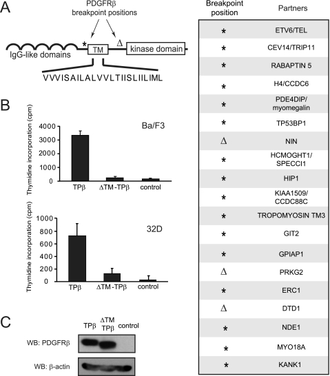
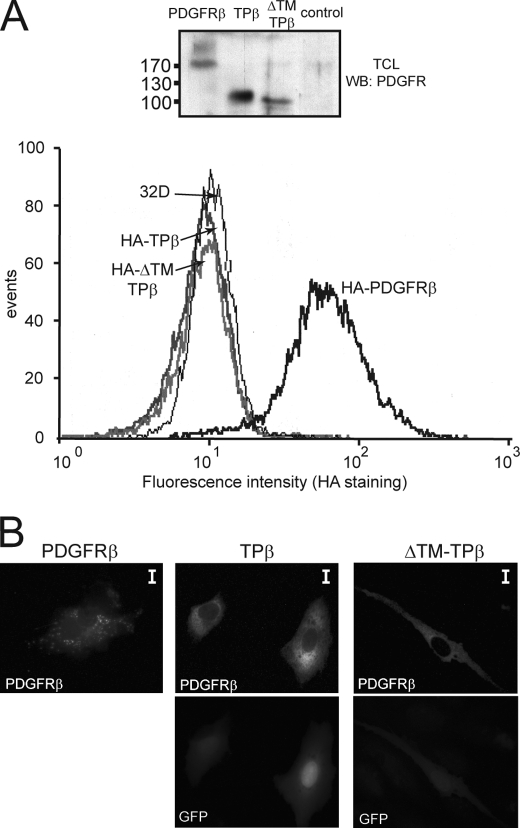
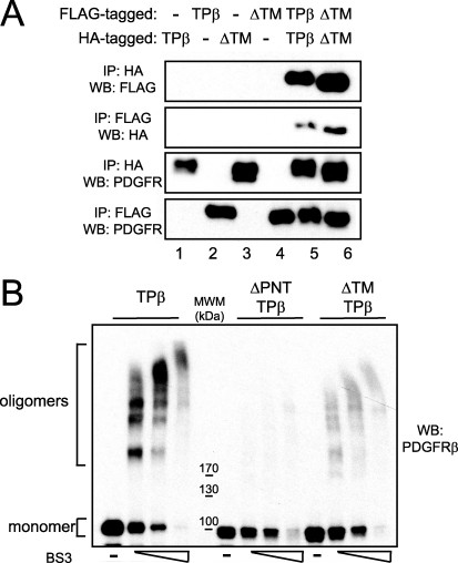
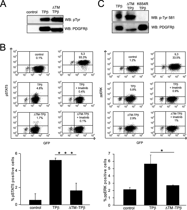
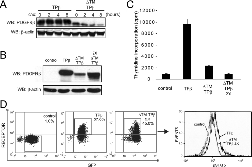
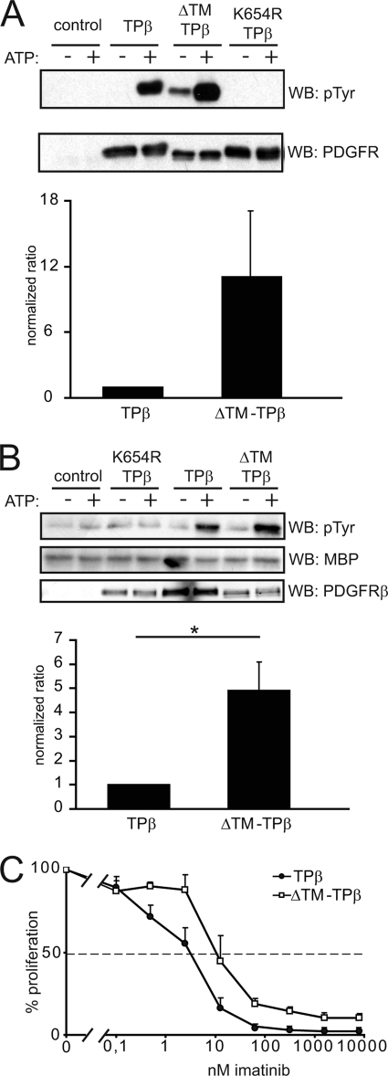
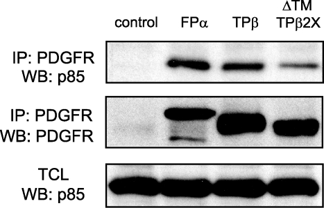
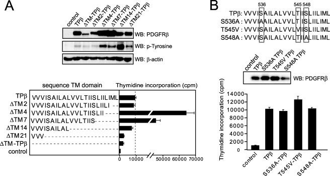
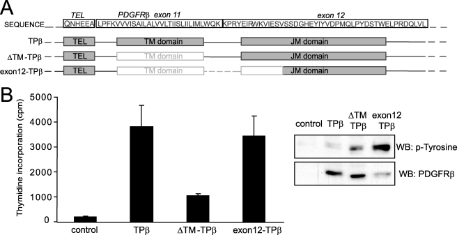
The fusion of TEL with platelet-derived growth factor receptor (PDGFR) beta (TPbeta) is found in a subset of patients with atypical myeloid neoplasms associated with eosinophilia and is the archetype of a larger group of hybrid receptors that are produced by rearrangements of PDGFR genes. TPbeta is activated by oligomerization mediated by the pointed domain of TEL/ETV6, leading to constitutive activation of the PDGFRbeta kinase domain. The receptor transmembrane (TM) domain is retained in TPbeta and in most of the described PDGFRbeta hybrids. Deletion of the TM domain (DeltaTM-TPbeta) strongly impaired the ability of TPbeta to sustain growth factor-independent cell proliferation. We confirmed that TPbeta resides in the cytosol, indicating that the PDGFRbeta TM domain does not act as a transmembrane domain in the context of the hybrid receptor but has a completely different function. The DeltaTM-TPbeta protein was expressed at a lower level because of increased degradation. It could form oligomers, was phosphorylated at a slightly higher level, co-immunoprecipitated with the p85 adaptor protein, but showed a much reduced capacity to activate STAT5 and ERK1/2 in Ba/F3 cells, compared with TPbeta. In an in vitro kinase assay, DeltaTM-TPbeta was more active than TPbeta and less sensitive to imatinib, a PDGFR inhibitor. In conclusion, we show that the TM domain is required for TPbeta-mediated signaling and proliferation, suggesting that the activation of the PDGFRbeta kinase domain is not enough for cell transformation.


Figures

FIGURE 1.
FIGURE 1.
The PDGFRβ transmembrane domain is critical for TPβ-mediated proliferation of hematopoietic cells. A, the domain organization of PDGFRβ is shown with the two main breakpoint positions depicted with an asterisk and an open triangle. The position marked by an asterisk is usually in the intron preceding exon 11, which encodes the TM sequence, but breakpoints in introns before exon 9 or 10 were also reported. The sequence of the TM domain is shown in capital letters. In the right panel are listed the fusion partners of PDGFRβ with the corresponding breakpoint positions in PDGFRβ. B, Ba/F3 or 32D cell lines were transduced with TPβ or ΔTM-TPβ using a bicistronic retroviral vector encoding GFP and sorted according to GFP levels. The cells were incubated for 24 h in absence of IL3. Proliferation was measured by a [3H]thymidine incorporation assay as described under “Experimental Procedures.” All of the cell lines proliferated equally in the presence of IL3 (data not shown). S.D. were calculated from triplicate cultures in a representative experiment. Vector-transfected cells were used as a control. C, total cell lysates derived from the Ba/F3 cell lines used in B were analyzed by Western blot (WB) with anti-PDGFR and anti-β-actin antibodies. The total cell lysates derived from the 32D cell lines were analyzed similarly (shown in Fig. 2A).
FIGURE 2.
FIGURE 2.
TPβ is not a membrane spanning protein. A, intact 32D cells stably expressing the HA-tagged form of wild-type PDGFRβ, TPβ, or ΔTM-TPβ were stained with anti-HA antibodies and analyzed by flow cytometry. Untransfected 32D cells were used as control. Total cell lysates (TCL) from the same cell lines were analyzed by Western blot (WB) with anti-PDGFRβ antibodies. B, PAE cells were transfected with the indicated receptors and stained with anti-PDGFRβ antibodies and fluorescent secondary antibodies. The cells were analyzed by fluorescent microscopy. GFP is co-expressed with TPβ and ΔTM-TPβ from the bicistronic vector used for transfection. The scale bars correspond to 10 μm.
FIGURE 3.
FIGURE 3.
ΔTM-TPβ oligomerization. A, 293T cells were transfected with the HA- or FLAG-tagged forms of TPβ or ΔTM-TPβ as indicated. Hybrid receptors were immunoprecipitated (IP) with anti-HA or anti-FLAG antibodies and then immunoblotted with anti-HA, anti-FLAG, or anti-PDGFRβ antibodies as indicated. B, cell lysates obtained from Ba/F3 cell lines expressing TPβ, ΔTM-TPβ, or ΔPNT-TPβ were left untreated or treated with increasing concentrations of BS3 as described under “Experimental Procedures.” The cell lysates were analyzed for the presence of oligomers by immunoblotting with anti-PDGFRβ antibodies. MWM, molecular weight marker. WB, Western blot.
FIGURE 4.
FIGURE 4.
ΔTM-TPβ is phosphorylated but showed impaired STAT5 and ERK1/2 activation. A, total cell lysates from Ba/F3 cells expressing the indicated receptors were analyzed for receptor phosphorylation by Western blot (WB) with anti-phosphotyrosine antibodies and then reblotted with anti-PDGFRβ antibodies. Vector-transfected cells were used as control. B, Ba/F3 cells expressing the indicated hybrid receptors were starved for 4 h. The cells were then permeabilized and stained with anti-pSTAT5 or anti-ppERK antibodies and analyzed by flow cytometry as described under “Experimental Procedures.” As controls, the cells were treated with IL3 for 5 min or with imatinib for 4 h before the fixation step. The percentages of GFP and pSTAT5/ppERK-positive cells are indicated in the upper right quadrant of the dot plots shown. The averages of at least two independent experiments are shown in the histogram with S.D. *, p ≤ 0.05; ***, p ≤ 0.001. C, total cell lysates from TPβ or ΔTM-TPβ were analyzed by Western blot with anti-PDGFRβ phosphotyrosine 581 antibodies. The kinase inactive mutant K654R-TPβ was used as negative control for the specificity of the phosphotyrosine antibody. The filter was stripped and reblotted with anti-PDGFRβ antibodies.
FIGURE 5.
FIGURE 5.
The ΔTM-TPβ protein is degraded faster. A, Ba/F3 cells expressing the indicated forms of hybrid receptors were used in stability assays with 50 μg/ml cycloheximide as described under “Experimental Procedures.” The samples were collected at different time points and analyzed by Western blot (WB) with anti-PDGFRβ and anti-β-actin antibodies. B, total cell lysates from Ba/F3 cells expressing TPβ, ΔTM-TPβ transduced only once with retroviruses (ΔTM-TPβ), or those transduced twice (ΔTM-TPβ2X) were analyzed with anti-PDGFRβ and anti-β-actin antibodies. Vector-transduced cells were used as a control. All of the samples were analyzed on the same Western blot, and some lanes were cut out of the final image for clarity. C, the same cell lines as in B were used in a proliferation assay as described for Fig. 1B. All of the cell lines proliferated equally in the presence of IL3 (data not shown). The cells transduced once or twice with TPβ proliferated at a similar level (data not shown). D, TPβ and ΔTM-TPβ2X cells were stained for phospho-STAT5 and PDGFRβ. The dot plots show the percentages of cells expressing GFP and PDGFRβ. The histogram shows the phospho-STAT5 staining in the population positive for GFP and for the PDGFRβ indicated by rectangles. TPβ is shown with a bold line, ΔTM-TPβ is shown with a thin line, and control is shown with a dashed line. One representative experiment is shown (n = 4). Vector-expressing cells were used as a control.
FIGURE 6.
FIGURE 6.
Deletion of the TM domain increases TPβ kinase activity in vitro. A, 293T cells were transfected with the indicated forms of the receptors and treated with 1 μm imatinib for 4 h to prevent receptor phosphorylation. Hybrid receptors were then immunoprecipitated with anti-PDGFRβ antibodies, extensively washed to remove imatinib, and used in an in vitro phosphorylation assay in the presence or absence of ATP. The reaction products were analyzed by immunoblotting with anti-phosphotyrosine and anti-PDGFRβ antibodies. The blots were quantified using an Odyssey instrument. Phosphorylation data were normalized by dividing by the total amount of receptor after background substraction (values in the absence of ATP). The results from two independent experiments were expressed as average fold increase compared with TPβ. B, receptors were expressed in cells and treated as in A except that the imatinib treatment was not applied. The in vitro reactions were performed in the presence of 10 μg of MBP. The reaction products were analyzed by anti-phosphotyrosine and anti-MBP immunoblotting. MBP phosphorylation values were normalized by dividing by the total amount of receptor, after background subtraction. The results from two independent experiments were expressed as average fold increase compared with TPβ. C, Ba/F3 cells expressing TPβ or ΔTM-TPβ were used in [3H]thymidine incorporation assays in the presence of increasing doses of imatinib. Proliferation was expressed as a percentage of the condition without imatinib. WB, Western blot.
FIGURE 7.
FIGURE 7.
ΔTM-TPβ can associate with p85. The indicated receptors were immunoprecipitated (IP) from stably expressing Ba/F3 cell lines and immunoblotted with anti-p85 and anti-PDGFR antibodies. Total cell lysates were analyzed with anti-p85 antibodies. FIP1L1-PDGFRα- (FPα) and vector-expressing cells were used as a control. WB, Western blot.
FIGURE 8.
FIGURE 8.
Transmembrane sequence requirements for optimal TPβ activation. A, the transmembrane sequence of each TPβ deletion mutant is shown. Mutant hybrids were transduced in Ba/F3 cells, and their ability to stimulate cell growth in the absence of cytokine was tested by thymidine incorporation as described for Fig. 1B. Vector-transfected cells were used as control. All of the cell lines proliferated equally in the presence of IL3 (data not shown). The total cell lysates were analyzed by Western blot (WB) with anti-phosphotyrosine, anti-PDGFRβ, and anti-β-actin antibodies. B, the same experiments were performed with the TPβ mutants S536A, T545V, and S548A.
FIGURE 9.
FIGURE 9.
Interruption of the juxtamembrane domain can overcome the effect of the deletion of the TM domain in TPβ. A, the positions of the TM and JM domains are indicated in the gray boxes below the amino acid sequence of TPβ. The deletions in ΔTM-TPβ and in exon12-TPβ are indicated by faded boxes. B, Ba/F3 cell lines expressing the indicated forms of hybrid receptors were used in a proliferation assay as described for Fig. 1B. The total cell lysates derived from the same cells were analyzed by Western blot (WB) with anti-PDGFRβ and anti-phosphotyrosine antibodies. All of the cell lines proliferated equally in the presence of IL3 (data not shown).

Similar articles

Cited by

References

    1. Andrae J., Gallini R., Betsholtz C. (2008) Genes Dev. 22, 1276–1312 - PMC - PubMed
    1. Schlessinger J. (2000) Cell 103, 211–225 - PubMed
    1. Yang Y., Yuzawa S., Schlessinger J. (2008) Proc. Natl. Acad. Sci. U.S.A. 105, 7681–7686 - PMC - PubMed
    1. Yuzawa S., Opatowsky Y., Zhang Z., Mandiyan V., Lax I., Schlessinger J. (2007) Cell 130, 323–334 - PubMed
    1. Chan P. M., Ilangumaran S., La Rose J., Chakrabartty A., Rottapel R. (2003) Mol. Cell. Biol. 23, 3067–3078 - PMC - PubMed

Publication types

MeSH terms

Substances