. 2010 Apr 9;285(15):11402-10.
doi: 10.1074/jbc.M110.105403. Epub 2010 Feb 17.

A novel protease-activated receptor-1 interactor, Bicaudal D1, regulates G protein signaling and internalization

Affiliations

A novel protease-activated receptor-1 interactor, Bicaudal D1, regulates G protein signaling and internalization

Steven Swift et al. J Biol Chem. .

Abstract

Protease-activated receptor-1 (PAR1) is a G protein-coupled receptor that plays critical roles in cancer, angiogenesis, inflammation, and thrombosis. Proteolytic cleavage of the extracellular domain of PAR1 generates a tethered ligand that activates PAR1 in an unusual intramolecular mode. The signal emanating from the irreversibly cleaved PAR1 is terminated by G protein uncoupling and internalization; however, the mechanisms of PAR1 signal shut off still remain unclear. Using a yeast two-hybrid screen, we identified Bicaudal D1 (BicD1) as a direct interactor with the C-terminal cytoplasmic domain of PAR1. BICD was originally identified as an essential developmental gene associated with mRNA and Golgi-endoplasmic reticulum transport. We discovered a novel function of BicD1 in the modulation of G protein signaling, cell proliferation, and endocytosis downstream of PAR1. BicD1 and its C-terminal CC3 domain inhibited PAR1 signaling to G(q)-phospholipase C-beta through coiled-coil interactions with the cytoplasmic 8th helix of PAR1. Unexpectedly, BicD1 was also found to be a potent suppressor of PAR1-driven proliferation of breast carcinoma cells. The growth-suppressing effects of BicD1 required the ability to interact with the 8th helix of PAR1. Silencing of BicD1 expression impaired endocytosis of PAR1, and BicD1 co-localized with PAR1 and tubulin, implicating BicD1 as an important adapter protein involved in the transport of PAR1 from the plasma membrane to endosomal vesicles. Together, these findings provide a link between PAR1 signal termination and internalization through the non-G protein effector, BicD1.


Figures
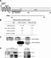
FIGURE 1.
FIGURE 1.
BicD1 associates with the C-terminal cytoplasmic domain of PAR1. A, model depicting the C-terminal cytoplasmic domains of PAR1 and PAR4 is shown. B, yeast two-hybrid screen of a mouse embryo cDNA library was conducted using GAL4DB-PAR1(373–425) or GAL4DB-PAR4(344–385) as the bait proteins. C, C-terminal CC3 domain of mBicD1, BicD1ct, was found to interact with GAL4DB-PAR1(373–425). Full-length BicD1 has three coiled-coil domains, CC1, CC2, and CC3. D, left immunoloblots, PAR1 was immunoprecipitated (IP) with the SFLLR antibody from COS7 cell lysates co-transfected with PAR1 and T7-BicD1, or pcDEF3 vector control. Right immunoblots, endogenous BicD1 was co-immunoprecipitated with the M2 FLAG antibody from Rat1 fibroblasts lysates stably transfected with FLAG-PAR1 and the blots stained with the BicD1 antibody or the SFLLR antibody, as indicated.
FIGURE 2.
FIGURE 2.
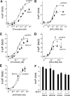
BicD1 specifically inhibits PAR1-G protein signaling. A–D, WT PAR1 was co-transfected into COS7 cells along with full-length BicD1(1–873), GFP-BicD1ct (BicD1ct), or pCDEF3 vector control as indicated. Cells were then challenged for 30 min with 1 pm to 10 nm thrombin, or 10 nm to 30 μm SFLLRN agonists (each point done in triplicate). PLC-β activity was determined by measuring total [3H]InsP formation and converted to fold response relative to buffer alone. Experiments were conducted at least three times. E, WT PAR4 was co-transfected into COS7 cells along with GFP-BicD1ct or pCDEF3 vector control and PLC-β activity was determined as above except that AYPGKF was used as agonist. F, full-length BicD1 (+) or pCDEF3 vector (−) was co-transfected into COS7 cells along with PAR2, SSTR2, CCKA, or CCKB receptors, and PLC-β activity was determined as above. The agonists used were 10 μm SLIGKV for PAR2, 1 μm AGCKNFFWKTFTSC for SSTR2, 300 nm CCK-8 for CCKA and CCKB. Data are plotted as mean ± 1 S.D. (error bars).
FIGURE 3.
FIGURE 3.
BicD1 inhibits G protein signaling through its interaction with the 8th helix of PAR1. A, C-terminal sequences of the PAR1 C-terminal domain and 8th helix mutants are shown. The two jagged lines over C387C388 represent putative palmitoylation sites. B–F, PAR1 mutants were co-transfected into COS7 cells along with GFP-BicD1ct (BicD1ct), full-length BicD1(1–873 aa), or pCDEF3 vector control as indicated. Cells were then challenged for 30 min with 1 pm to 100 nm thrombin (each point done in triplicate), and PLC-β activity was determined by measuring total [3H]InsP formation and converted to fold response relative to buffer alone (n = 3). Data are plotted as mean ± 1 S.D. (error bars). G, co-immunoprecipitation (IP) of BicD1 with PAR1 requires an intact 8th helix of PAR1. COS7 cells were transiently transfected with PAR1 mutants and full-length T7-BicD1. Lysates from transfected COS7 were incubated with T7-agarose beads and proteins in lysate, and immunoprecipitation eluates were separated by 10% SDS-PAGE, and Western blot analysis (IB) using the SFLLR and BicD1 antibodies were performed.
FIGURE 4.
FIGURE 4.
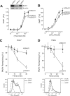
Knockdown of BicD1 expression enhances PAR1-G protein signaling and inhibits endocytosis. A and B, PAR1 was co-transfected into Rat1 cells along with antisense-BicD1 DNA, antisense-EGFP DNA, or pCDEF3 vector controls as indicated. Cells were then challenged for 30 min, or SFLLRN and PLC-β activity was determined by measuring total [3H]InsP formation and converted to percent response relative to the maximal signal observed with pCDEF3 control (n = 3). Knockdown of BicD1 by asBicD1 was confirmed by Western blot analysis as shown in the inset in A. C and D, upper, endocytosis of PAR1 in COS7 cells (C) and HeLa cells (D) co-transfected with PAR1 and antisense BicD1 or pCDEF3 control vector is indicated. Loss of PAR1 SFLLR epitope from the cell surface was analyzed by flow cytometry as described previously (3). Lower, basal PAR1 surface expression levels of both COS7 and HeLa cells determined by FACS indicate that the surface expression levels of PAR1 in the antisense BicD1-transfected cells are not significantly different from control pcDEF3 cells. Data are plotted as mean ± 1 S.D. (error bars). *, p < 0.05.
FIGURE 5.
FIGURE 5.
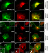
Subcellular localization of BicD1 and PAR1. COS7 cells were transfected with GFP-tubulin (A and B), PAR1 (C–E), and T7-BicD1 (A–E) and grown on coverslips. After 2 days, cells were fixed and stained with T7 antibody and SFLLRN antibody. A and B, epifluorescence (A) and confocal fluorescence microscopy (B) of untreated cells expressing BicD1 and GFP-tubulin. C, epifluorescence microscopy of untreated cells expressing PAR1 and BicD1. D and E, confocal microscopy of cells expressing BicD1 and PAR1 treated for 30 min with phosphate-buffered saline (D) or 30 μm SFLLRN (E).
FIGURE 6.
FIGURE 6.
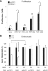
Knockdown of BicD1 expression or mutation of the BicD1-interacting region of PAR1 enhances proliferation and modulates ligand-dependent endocytosis in MCF-7 breast carcinoma cells. A, MCF-7 cells (2500/well) were transiently transfected with WT, ΔH8, SIL, or PAR1Δ396 along with pcDEF3 or asBicD1 in 96-well plates. After 48 h of transfection, cells were allowed to grow for another 72 h in RPMI 1640 serum-free medium, and proliferation was measured by 3-(4,5-dimethylthiazol-2-yl)-2,5-diphenyltetrazolium bromide assay (n = 4, mean ± S.D. (error bars)) as described previously (40). B, endocytosis of PAR1 in MCF-7 cells co-transfected with WT or SIL-PAR1 plus pcDEF3, BicD1, or asBicD1 is shown. Loss of PAR1 SFLLR-epitope from the cell surface was analyzed by flow cytometry as in Fig. 4. Data are plotted as mean ± 1 S.D. (error bars). *, p < 0.05; **, p < 0.001; ns, nonsignificant.

References

    1. Ossovskaya V. S., Bunnett N. W. (2004) Physiol. Rev. 84, 579–621 - PubMed
    1. Vu T.-K. H., Hung D. T., Wheaton V. I., Coughlin S. R. (1991) Cell 64, 1057–1068 - PubMed
    1. Kuliopulos A., Covic L., Seeley S. K., Sheridan P. J., Helin J., Costello C. E. (1999) Biochemistry 38, 4572–4585 - PubMed
    1. Riewald M., Petrovan R. J., Donner A., Mueller B. M., Ruf W. (2002) Science 296, 1880–1882 - PubMed
    1. Boire A., Covic L., Agarwal A., Jacques S., Sherifi S., Kuliopulos A. (2005) Cell 120, 303–313 - PubMed

Publication types

Substances