. 2010 Apr 16;285(16):12121-32.
doi: 10.1074/jbc.M109.075721. Epub 2010 Feb 17.

Interactions among HAMP domain repeats act as an osmosensing molecular switch in group III hybrid histidine kinases from fungi

Affiliations

Interactions among HAMP domain repeats act as an osmosensing molecular switch in group III hybrid histidine kinases from fungi

Netrapal Meena et al. J Biol Chem. .

Abstract

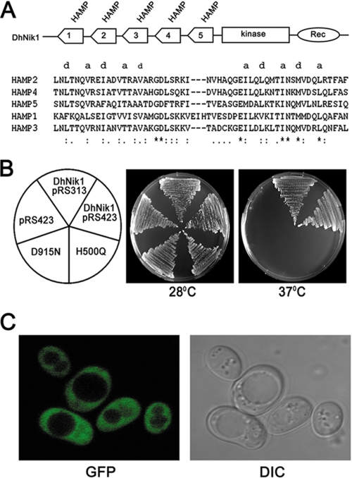
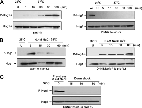
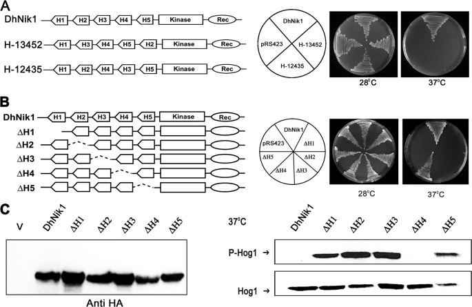
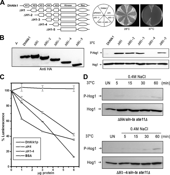
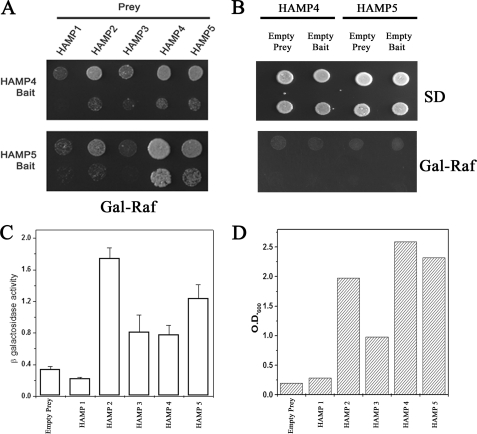
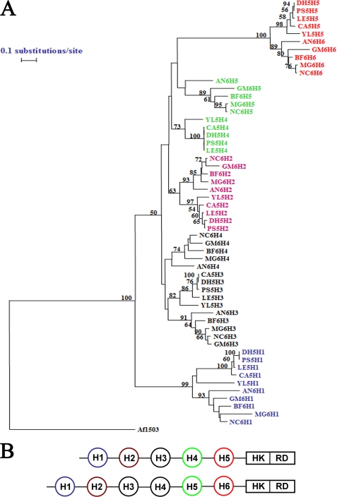
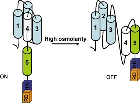
The members of group III hybrid histidine kinases (HHK) are ubiquitous in fungi. Group III HHK have been implicated to function as osmosensors in the high osmolarity glycerol (HOG) pathway that is essential for fungal survival under high osmolarity stress. Recent literature suggests that group III HHK are also involved in conidia formation, virulence in several filamentous fungi, and are an excellent molecular target for antifungal agents. Thus, group III HHK constitute a very important group of sensor kinases. Structurally, group III HHK are distinct from Sln1p, the osmosensing HHK that regulates the HOG pathway in Saccharomyces cerevisiae. Group III HHK lack any transmembrane domain and typically contain HAMP domain repeats at the N terminus. Until now, it is not clear how group III HHK function as an osmosensor to regulate the HOG pathway. To investigate this, we undertook molecular characterization of DhNIK1, an ortholog from osmotolerant yeast Debaryomyces hansenii. We show here that DhNIK1 could complement sln1 mutation in S. cerevisiae thereby confirming its role as a bona fide osmosensor. We further investigated the role of HAMP domains by deleting them systematically. Our results clearly indicate that the HAMP4 domain is crucial for osmosensing by DhNik1p. Most importantly, we also show that the alternative interaction among the HAMP domains regulates the activity of DhNik1p like an "on-off switch" and thus provides, for the first time, an insight into the molecular mechanism of osmosensing by this group of HHKs.


Figures

FIGURE 1.
FIGURE 1.
Complementation of sln1 mutation in S. cerevisiae by DhNIK1. A, diagrammatic representation of the domain architecture of DhNik1p. HAMP domains (numbered serially according to their position from the N terminus), histidine kinase (Kinase), and receiver (Rec) domains are shown. Multiple alignments are shown of amino acid residues of the individual HAMP domains using ClustalX1.8. Hydrophobic residues of the two amphipathic helices are denoted by letter a and d to show their heptad periodicity. B, growth of TM229 transformed with only vector pRS423, DhNIK1 in multicopy vector pRS423 or single copy vector pRS313, mutants of DhNIK1 H500Q or D915N on SD agar plate without histidine at 28 or 37 °C after 3 days of incubation. C, subcellular localization of DhNik1p by fluorescence microscopy. Strain TM229 expressing the DhNik1-GFP fusion protein was grown to logarithmic phase in SD minimal medium, and the GFP fluorescence was viewed under an LSM510 META laser scanning confocal microscope (Carl Zeiss). GFP fluorescence (GFP) and differential interference contrast (DIC) image of the same cells are shown.
FIGURE 2.
FIGURE 2.
Regulation of HOG pathway in S. cerevisiae by DhNIK1. A, immunoblots showing the level of Hog1p phosphorylation in S. cerevisiae strain TM229 (sln1-ts) or TM229 expressing DhNik1p at nonpermissive temperature (37 °C). Cells were grown on SD medium with the required nutrient supplement at 28 °C until exponential phase (A600 ∼0.7) before exposing them to 37 °C for indicated time. The level of dually phosphorylated Hog1p (P-Hog1) was detected by immunoblotting the total cell extracts with anti-phospho-p38 antibody (Cell Signaling Inc). The total extract from cells grown at 28 °C was loaded as control (U). The total cell extracts from TM229 exposed to 37 °C for 1 h are included in the right panel as positive control (+ve). Blots were re-probed with anti Hog1 antibody (Y215; Santa Cruz Biotechnology) to detect total Hog1. P-Hog1 or Hog1 indicate phosphorylated or total Hog1p in the blot. B, immunoblots showing high osmolarity-induced Hog1p phosphorylation in S. cerevisiae strain NM2 (sln1-ts ste11Δ) at 28 °C (left panel) and NM2 expressing DhNik1p at 37 °C (right panel). Cells at A600 ∼0.7 were exposed to 0.4 m NaCl for different lengths of time as indicated, and total cell extract from these samples was used for blotting. Total cell extracts from respective cultures not exposed to osmotic stress were loaded as control (U). C, level of dually phosphorylated Hog1p after down-shifting to low osmolarity. NM2 carrying DhNIK1 was grown at 28 °C up to A600 ∼0.7. Cells were incubated at 37 °C for another hour before exposing to 0.4 m NaCl for 5 min at 37 °C. Following centrifugation at 37 °C (to prevent the reactivation of sln1-ts), the cell pellet was resuspended in SD media without salt (maintained at 37 °C) and incubated further at 37 °C for different times as indicated. Total protein extract made from these samples were immunoblotted. All the blots shown above are representative of at least three different experiments.
FIGURE 3.
FIGURE 3.
Functional analysis of reshuffled and individually deleted HAMP domain mutants of DhNik1p. A, diagrammatic representations of DhNik1, HAMP domain reshuffled mutants H13452, or H12435 are shown in the left panel. HAMP domains are named H1, H2, H3, H4, and H5 according to their positions in DhNik1. To determine the growth patterns of TM229 harboring vector (pRS423), DhNik1, H13452, or H12435, cells were streaked on minimal SD agar plate without histidine and incubated at 28 or 37 °C for 3 days. Photographs of the respective plates are shown. B, diagrammatic representations of single HAMP domain deletion mutants and their growth patterns on minimal SD agar plate without histidine at 28 or 37 °C are shown. Experiments were repeated three times with similar results. C, left panel, immunoblot showing the level of expression of DhNik1p and HAMP-deleted mutant ΔH1, ΔH2, ΔH3, ΔH4, and ΔH5. Total cell extracts from TM229 expressing HA-tagged DhNik1 and different mutants were immunoblotted with anti-HA antibody (Cell Signaling). Total cell extract from TM229 transformed with pRS423 were loaded as control (V). Right panel, immunoblots showing the phosphorylated Hog1p in cells with different HAMP domain deletion mutants. S. cerevisiae strain TM229 (sln1-ts) carrying DhNik1 or different mutants (ΔH1, ΔH2, ΔH3, ΔH4, and ΔH5) were grown on SD minimal media without histidine at 28 °C until an A600 ∼0.7 and exposed to 37 °C for 1 h. Total protein extract from these cells was analyzed by Western blotting. P-Hog1 or Hog1 indicate phosphorylated or total Hog1p in the blot. Representative blots of three different experiments are shown.
FIGURE 4.
FIGURE 4.
Effect of serial deletions of HAMP domains in DhNIK1. A, diagrammatic representations of serially deleted HAMP domain mutants and growth patterns of TM229 carrying vector pRS423, DhNik1, or different HAMP domain deleted mutants on minimal SD agar plate without histidine at 28 or 37 °C are shown. Experiments were repeated three times with similar results. B, left panel, immunoblot showing the level of expression of DhNik1p, HAMP-deleted mutants ΔH1, ΔH1–2, ΔH1–3, ΔH1–4, and ΔH1–5 after expressing them as HA-tagged proteins. Total cell extract from TM229 transformed with pRS423 were loaded as control (V). Right panel, immunoblots showing the phosphorylated Hog1p in cells with different HAMP domain deletion mutants. S. cerevisiae strain TM229 (sln1-ts) carrying DhNik1 or different mutants (ΔH1, ΔH1–2, ΔH1–3, ΔH1–4, and ΔH1–5) were grown on SD minimal media without histidine at 28 °C until A600 ∼0.7 and exposed to 37 °C for 1 h. The level of phosphorylated Hog1p in these cells was detected by immunoblotting as described previously. Experiments were repeated three times with similar results. C, kinase activities of DhNik1p, ΔH4, and ΔH1–4 were measured by a luminescence assay. Assay was done without any protein or with 1, 3, and 6 μg of purified, recombinant proteins (DhNik1p, ΔH4, and ΔH1–4). Decreasing luminescence indicated increasing kinase activity. Bovine serum albumin (BSA) was used as a negative control. The luminescence observed with different protein concentrations was expressed as percentage of that obtained with the corresponding reaction without any protein. Data are the means ± S.D. of three experiments. D, high osmolarity induced Hog1p phosphorylation in S. cerevisiae strain NM2 (sln1-ts ste11Δ) expressing HAMP domain deletion mutant ΔH4 and mutant ΔH1–4. Cells were grown on SD minimal media without histidine at 28 °C until A600 ∼0.7 and exposed to 37 °C for 1 h. Osmotic stress was given to these cells by exposing them to 0.4 m NaCl at 37 °C for different lengths of time as indicated, and total cell extract from these samples was used for blotting. Total cell extract from respective culture not exposed to osmostress (Un) was loaded as control. Blots were re-probed with anti Hog1 antibody. P-Hog1 or Hog1 indicate phosphorylated or total Hog1p in the blot.
FIGURE 5.
FIGURE 5.
Interactions between HAMP domains of DhNik1p. A, two-hybrid assay for interactions between HAMP domains. HAMP4 or HAMP5 cloned in pEG202 vector was used as bait, and five HAMP domains were cloned individually in plasmid pJG4-5 for prey construction. Both bait and prey constructs (in pairs as indicated) were transformed into S. cerevisiae strain EGY48. Growth of the transformants on Gal-Raf minimal medium is shown after dilution spotting. Results are representative of three different experiments. B, two-hybrid interactions in HAMP4 and HAMP5 with empty bait or prey combinations. S. cerevisiae strain EGY48 transformed with HAMP4 bait/empty prey; HAMP4 prey/empty bait; HAMP5 bait/empty prey, and HAMP5 prey/empty bait were grown in minimal SD medium, and serial dilution of the culture was spotted onto minimal SD and Gal-Raf plate. C, β-galactosidase activity in yeast strain EGY48 harboring lacZ reporter plasmid, HAMP4 bait along with empty prey or different HAMP domains in prey vector. Experiments were repeated with two independent pools of six transformants each. β-Galactosidase activity is expressed as nanomoles of o-nitrophenyl β-d-galactopyranoside utilized per min by 1 ml of culture after normalizing its A600 to 1.0. D, EGY48 harboring HAMP4 bait along with empty prey or different HAMP domains in prey vector were grown overnight in minimal media with 2% raffinose (without tryptophan and histidine). The cultures were re-inoculated in minimal media with 1% raffinose and 2% galactose (without tryptophan, histidine and leucine) at A600 ∼0.10 and grown further for 40 h. Representative data of two independent experiments are shown here.
FIGURE 6.
FIGURE 6.
Phylogenetic analysis of HAMP domains. A, phylogenetic tree. Individual HAMP domain sequences from the NIK1 orthologs having five HAMP domains from Yarrowia lipolytica (Q6C775_YARLI), Lodderomyces elongisporus (A5E5X7_LODEL), P. stipitis (A3LYT9_PICST), D. hansenii (DhNik1p; Q6BH10_DEBHA), and C. albicans (CaNik1p; Q9URL9_CANAL) were obtained from SMART data base. YL5H1, YL5H2, YL5H3, YL5H4, and YL5H5 were HAMP domains from Y. lipolytica ortholog. They were named serially according to their position in the protein from N terminus. Similarly, LE5H1, LE5H2, LE5H3, LE5H4, and LE5H5 were from L. elongisporus; PS5H1, PS5H2, PS5H3, PS5H4, and PS5H5 were from P. stipitis; DH5H1, DH5H2, DH5H3, DH5H4, and DH5H5 were from D. hansenii; CA5H1, CA5H2, CA5H3, CA5H4, and CA5H5 were from C. albicans. Representative orthologs of NIK1 containing six HAMP domains are B. fuckeliana (Q8X1E7_BOTCI), Gibberella moniliformis (Q6SLB2_GIBMO), N. crassa (Nik1p; Q01309_NEUCR), M. grisea (Q9C1U1_MAGGR), and Aspergillus niger (A2QP39_ASPNG). BF6H1, BF6H2, BF6H3, BF6H4, BF6H5, and BF6H6 are individual HAMP domain sequences (named serially according to their position from N terminus) from ortholog in B. fuckeliana. Similarly, GM6H1, GM6H2, GM6H3, GM6H4, GM6H5, and GM6H6 were from G. moniliformis; NC6H1, NC6H2, NC6H3, NC6H4, NC6H5, and NC6H6 were from N. crassa; MG6H1, MG6H2, MG6H3, MG6H4, MG6H5, and MG6H6 were from M. grisea; AN6H1, AN6H2, AN6H3, AN6H4, AN6H5, and AN6H6 were from A. niger. Amino acid sequences of the individual HAMP domains were compared using the Clustal_X program (supplemental Table 2 and supplemental Fig. 1). Aligned sequences were analyzed by using the TREECON software package. A neighbor-joining tree (50% or more boot strap values of 1000 replicates are indicated) is shown. HAMP domain sequence from Archaeoglobus fulgidus protein Af1503 (30) was included as out-group. B, schematic showing Nik1 orthologs carrying five HAMP domains and six HAMP domains. Similarly colored HAMP domains from either group are clustered together in the phylogenetic tree. HK, histidine kinase; RD, receiver domain.
FIGURE 7.
FIGURE 7.
Model depicting how the activity of DhNik1 is regulated under high and low osmolarity conditions. Each of the HAMP domains (cylinder) is numbered. The histidine kinase and receiver domain are shown in box labeled as HK and RD. Under high osmolarity stress, HAMP4 interacts with HAMP5 to shut off the kinase activity of DhNik1p.

Comment in

Similar articles

Cited by

References

    1. Yamada-Okabe T., Mio T., Ono N., Kashima Y., Matsui M., Arisawa M., Yamada-Okabe H. (1999) J. Bacteriol. 181, 7243–7247 - PMC - PubMed
    1. Santos J. L., Shiozaki K. (2001) Sci. STKE 2001, RE1. - PubMed
    1. Nemecek J. C., Wüthrich M., Klein B. S. (2006) Science 312, 583–588 - PubMed
    1. Viaud M., Fillinger S., Liu W., Polepalli J. S., Le Pêcheur P., Kunduru A. R., Leroux P., Legendre L. (2006) Mol. Plant-Microbe Interact. 19, 1042–1050 - PubMed
    1. Bahn Y. S., Xue C., Idnurm A., Rutherford J. C., Heitman J., Cardenas M. E. (2007) Nat. Rev. Microbiol. 5, 57–69 - PubMed

Publication types

MeSH terms