. 2010 Apr 9;285(15):11357-68.
doi: 10.1074/jbc.M110.102459. Epub 2010 Feb 17.

Hantavirus nucleocapsid protein has distinct m7G cap- and RNA-binding sites

Affiliations

Hantavirus nucleocapsid protein has distinct m7G cap- and RNA-binding sites

Mohammad A Mir et al. J Biol Chem. .

Abstract

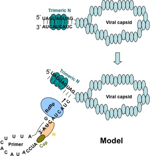
Hantaviruses, members of the Bunyaviridae family, are emerging category A pathogens that carry three negative stranded RNA molecules as their genome. Hantavirus nucleocapsid protein (N) is encoded by the smallest S segment genomic RNA (viral RNA). N specifically binds mRNA caps and requires four nucleotides adjacent to the cap for high affinity binding. We show that the N peptide has distinct cap- and RNA-binding sites that independently interact with mRNA cap and viral genomic RNA, respectively. In addition, N can simultaneously bind with both mRNA cap and vRNA. N undergoes distinct conformational changes after binding with either mRNA cap or vRNA or both mRNA cap and vRNA simultaneously. Hantavirus RNA-dependent RNA polymerase (RdRp) uses a capped RNA primer for transcription initiation. The capped RNA primer is generated from host cell mRNA by the cap-snatching mechanism and is supposed to anneal with the 3' terminus of vRNA template during transcription initiation by single G-C base pairing. We show that the capped RNA primer binds at the cap-binding site and induces a conformational change in N. The conformationally altered N with a capped primer loaded at the cap-binding site specifically binds the conserved 3' nine nucleotides of vRNA and assists the bound primer to anneal at the 3' terminus. We suggest that the cap-binding site of N, in conjunction with RdRp, plays a key role during the transcription and replication initiation of vRNA genome.


Figures

FIGURE 1.
FIGURE 1.
Expression and purification of wild type and mutant N protein. Bacterial lysate containing His-tagged wild type N (lane 1) was loaded onto a nickel-nitrilotriacetic acid column for purification. The flow through the column is shown in lane 2. After washing the column, bound N was eluted by running an imidazole gradient from 40–500 mm. Fractions containing N were pooled (lane 3). Pooled fractions were dialyzed and further purified on a size exclusion column (lane 4). Mutant N was processed similarly, and the final purified fraction of the mutant is shown in lane 6. The size of purified proteins matches correctly with the molecular weight marker (lane 5).
FIGURE 2.
FIGURE 2.
Fluorescence binding of m7GDP with N protein. A shows the fluorescence spectrum of N protein (150 nm) from 300–500 nm (top spectra) in RNA-binding buffer. Shown is the differential fluorescence spectrum of N, after incubation with 90 nm (middle) and 40 nm (bottom) m7GDP. A fluorescence signal of 40 and 90 nm m7GDP without N was subtracted. Fluorescence intensity at 330 nm was recorded at each input concentration of m7GDP. A plot of fluorescence intensity at 330 nm versus input m7GDP concentration is shown in B. ΔFFmax was calculated as described under “Experimental Procedures” and plotted against m7G concentration (C). Data points were fitted according to Equations 1 and 2 (under “Experimental Procedures”) for the calculation of Kd. a.u., arbitrary units.
FIGURE 3.
FIGURE 3.
Filter binding of different radiolabeled RNA molecules with N. A, results of a filter binding assay for the interaction of radiolabeled capped (filled squares) and uncapped (open circles) RNA decamer with N. Radiolabeled RNA decamers were incubated with increasing concentrations of N at room temperature and filtered through a nitrocellulose filter. The percentage of hot RNA retained on the filter is plotted versus increasing input concentration of N to generate the binding profile for the calculation of dissociation constants (shown in Table 2). Similarly, the binding profiles for the interaction of wild type N (open squares) and mutant N (filled squares) with SNV S segment RNA and capped RNA decamer, respectively, were generated (B). The binding profile for N-capped decamer interaction is replotted for comparison. C, in a competition assay, a fixed concentration of N (530 nm) was incubated with 0.01 nm radiolabeled capped decamer at increasing input concentrations of either cold capped decamer (filled squares) or cold viral S segment RNA (open squares) and filtered through a nitrocellulose filter. The percentage of hot capped decamer retained on the filter is plotted versus competitor RNA concentration. D, a filter binding assay was carried out in which a fixed concentration of radiolabeled SNV S segment was incubated with increasing concentrations of N-cap complex. The N-cap complex was generated by incubating N with saturating concentrations of cold capped RNA decamer. (see “Experimental Procedures” for details). Reaction mixtures were filtered through a nitrocellulose filter, and the percentage of hot S segment RNA retained on the filter is plotted versus input concentrations N-cap complex to generate the binding profile for the calculation of Kd. a.u., arbitrary units.
FIGURE 4.
FIGURE 4.
Stern-Volmer plots of N under different conditions. Stern-Volmer plots were generated by quenching the tryptophan fluorescence of N with acrylamide. A fixed concentration of N in RNA-binding buffer was excited at 295 nm, and the emission was recorded at 330 nm. The fluorescence studies were carried out at room temperature. The fluorescence signal from free binding buffer in absence of N was subtracted whenever required. The fluorescence intensity at 330 nm was determined at different input concentrations of acrylamide. Plots of F0/F as a function of acrylamide concentration (Stern-Volmer plot) for free N protein is shown by filled square in all three panels. Open squares show the Stern-Volmer plot of N bound to viral S segment RNA (A), capped decamer (B), and both the capped decamer and S segment vRNA simultaneously (C).
FIGURE 5.
FIGURE 5.
Fluorescence titration of N with hydrophobic fluorophore (bis-ANS) in RNA-binding buffer at room temperature under different conditions. The fluorophore was excited at 399 nm, and emission was recorded at 485 nm. Shown is the titration curve of bis-ANS binding with either free N (filled squares) or N that was preincubated with either S segment RNA (filled circles) or capped RNA decamer (filled triangles) or simultaneously with both capped decamer and S segment vRNA (open squares). For details, see under “Experimental Procedures.”
FIGURE 6.
FIGURE 6.
Sequence of primer-RNA pairs used in reverse transcription reaction (Fig. 7) are shown in panels A–L. Capped RNA primers had either terminal three (A) or a single (B) nucleotide complementary with the 3′ terminus of SNV S segment RNA. C and D are the same as A and B, respectively, except the primers used were uncapped. E and F are the same as A and B, respectively, except the sequence at the 3′ terminus was randomized in italic. G and H are same as A and B, respectively, except the template was a nonviral RNA of the same length as the SNV S segment and contained 3′ terminal nine nucleotides from the 3′ terminus of S segment. I and J are the same as A and B, respectively, except the vRNA template has three nucleotides deleted at the 3′ terminus. K and L are the same as A and B, respectively, except that five extra nucleotides are added to the 3′ terminus of the vRNA template.
FIGURE 7.
FIGURE 7.
Reverse transcription reactions. Reverse transcription reactions using primer-template pairs (A–L) from Fig. 6 were carried out as described in detail under “Experimental Procedures.” Reverse transcription products were radiolabeled during synthesis and run on 12% SDS-PAGE. The primer-template pairs used in reverse transcription reactions are shown at the top of the gel. In lanes 1 and 2, the primer and template were heated at 90 °C followed by cooling at room temperature to allow the annealing of the primer with the template. In lanes 3–18, the primer and template were incubated at room temperature without heating and cooling steps. All lanes from 5 to 18 (except lanes 9 and 10) contained wild type N. Lanes 9 and 10 contained mutant N that lacked the RNA-binding domain.
FIGURE 8.
FIGURE 8.
Filter binding assay showing the interaction of N with 3′ terminal nine nucleotides of SNV S segment RNA. Binding reactions were carried out as described in detail under “Experimental Procedures.” A shows a representative binding profile of N with a 3′ terminal nine-nucleotide-long triplet repeat sequence (5′-CUACUACUA-3′) of SNV S segment vRNA. The binding reaction was carried out in RNA-binding buffer containing 80 mm NaCl. In B, N protein was preincubated with saturating concentrations of cold capped RNA primer (shown in Fig. 6A) to generate the N-cap complex. The increasing concentrations of the complex were incubated with radiolabeled triplet repeat sequence and filtered through nitrocellulose filter. Radioactive signal retained on the filter at different input concentrations was used to generate the binding profile for the calculation of dissociation constants, shown in Table 3.
FIGURE 9.
FIGURE 9.
Model showing the role of N in primer annealing during transcription initiation. Hantaviral RNA, enveloped with N in viral capsids, is used as a template by viral RdRp during transcription and replication. Trimeric N binds the panhandle that is formed by the base pairing of complementary nucleotides at the 5′ and 3′ termini of vRNA. After binding, N unwinds the panhandle and remains attached with the 5′ terminus and leaves 3′ terminus accessible for RdRp. N with a capped RNA primer loaded at its cap binding site specifically recognizes the 3′ terminus of vRNA and assists the annealing of the bound primer.

Similar articles

Cited by

References

    1. Schmaljohn C. M. (1996) Molecular Biology of Hantaviruses, pp. 1–20, Plenum Press, New York
    1. Schmaljohn C. S., Hooper J. W. (2001) in Fields Virology (Howley K. A. ed.) pp. 1581–1602, Lippencott, Williams, and Wilkins, Philadelphia:
    1. Schmaljohn C. S., Jonsson C. B. (2001) in Hantaviruses (Nichol S. A. ed) pp. 15–32, Springer-Verlag, Berlin
    1. Obijeski J. F., Bishop D. H., Murphy F. A., Palmer E. L. (1976) J. Virol. 19, 985–997 - PMC - PubMed
    1. Pettersson R. F., von Bonsdorff C. H. (1975) J. Virol. 15, 386–392 - PMC - PubMed

Publication types