. 2010 Apr 16;285(16):12426-34.
doi: 10.1074/jbc.M109.068304. Epub 2010 Feb 17.

The phospholipid-binding protein SESTD1 is a novel regulator of the transient receptor potential channels TRPC4 and TRPC5

Affiliations

The phospholipid-binding protein SESTD1 is a novel regulator of the transient receptor potential channels TRPC4 and TRPC5

Susanne Miehe et al. J Biol Chem. .

Abstract

TRPC4 and TRPC5 are two closely related members of the mammalian transient receptor potential cation channel family that have been implicated in important physiological functions, such as growth cone guidance and smooth muscle contraction. To further unravel the role of TRPC4 and TRPC5 in these processes in vivo, detailed information about the molecular composition of native channel complexes and their association with cellular signaling networks is needed. We therefore searched a human aortic cDNA library for novel TRPC4-interacting proteins using a modified yeast two-hybrid assay. This screen identified SESTD1, a previously uncharacterized protein containing a lipid-binding SEC14-like domain as well as spectrin-type cytoskeleton interaction domains. SESTD1 was found to associate with TRPC4 and TRPC5 via the channel's calmodulin- and inositol 1,4,5-trisphosphate receptor-binding domain. In functional studies, we demonstrate that SESTD1 binds several phospholipid species in vitro and is essential for efficient receptor-mediated activation of TRPC5. Notably, phospholipid binding to SESTD1 was Ca(2+)-dependent. Because TRPC4 and -5 conduct Ca(2+), SESTD1-channel signaling may be bidirectional and also couple TRPC activity to lipid signaling through SESTD1. The modulation of TRPC channel function by specific lipid-binding proteins, such as SESTD1, adds another facet to the complex regulation of these channels complementary to the previously described effects of direct channel-phospholipid interaction.


Figures

FIGURE 1.
FIGURE 1.
Mapping of the SESTD1-binding site on the mTRPC4α C terminus. A, illustration of truncation mutants of the mTRPC4α C terminus used in yeast two-hybrid assays to identify the SESTD1-binding site. B, yeast colonies co-transformed with the given truncation mutants as baits and full-length human SESTD1 as prey were plated on selective −Trp/−Leu/−Ade/−His agar plates. Growth (light color) indicates protein-protein interaction. C, alignment of the identified SESTD1 binding site in mTRPC4α (amino acids (aa) 700–728) with mTRPC5 shows that this region is highly conserved between the two proteins (identical and diverse amino acids are denoted by the red and blue bars, respectively). The binding site of mTRPC4α is completely conserved in mTRPC4β. wt, wild type.
FIGURE 2.
FIGURE 2.
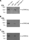
Binding of the SESTD1 spectrin 1 domain to TRPC4 and TRPC5. GST fusion proteins of the spectrin 1 or 2 domains of SESTD1 (GST-Spec1 or -2) were used to pull down full-length mTRPC4α (A), mTRPC4β (B), or mTRPC5 (C) from lysates of HEK293 cells transiently transfected with the respective cDNAs. Immunoprecipitates were Western blotted and stained with anti-TRPC4 (A and B) or anti-TRPC5 (C). 10% of the lysate input was run as expression control. GST alone was used to verify specificity of the pull-down assays.
FIGURE 3.
FIGURE 3.
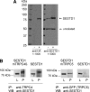
SESTD1 co-immunoprecipitates with mTRPC4β and mTRPC5. A, lysates of nontransfected HM1 cells (−) and cells transfected with HA-SESTD1 (+) were analyzed on Western blots using anti-HA antibodies and a polyclonal antibody against SESTD1. Both antibodies detected an ∼80-kDa protein in transfected cells, indicating that this protein is SESTD1. Anti-SESTD1 also detected an 80-kDa protein in nontransfected HM1 cells that we suggest is native SESTD1 (see also Fig. 7). In addition, the antibody recognized an unrelated ∼50-kDa protein. B, Western blot of anti-TRPC4 immunoprecipitates (P) and the corresponding lysates (L) from HM1 cells transfected with HA-tagged SESTD1 and FLAG-tagged mTRPC4β or HA-tagged SESTD1 alone (left). Shown are Western blots of anti-GFP immunoprecipitates (P) and the corresponding lysates (L) from HM1 cells transfected with HA-tagged SESTD1 and GFP-tagged mTRPC5 or HA-tagged SESTD1 alone (right). IP, immunoprecipitation; WB, Western blot.
FIGURE 4.
FIGURE 4.
Quantitative expression profiling of SESTD1 mRNA. SESTD1 mRNA expression was determined in different human tissues (top) or cell lines and selected primary cell types (bottom) using quantitative RT-PCR. Expression levels were normalized to the expression of the housekeeping gene RPL37a. Data shown are means of duplicates. Cell line designations are according to the ATCC and standard literature. Primary cells used were chondrocytes, peripheral blood mononuclear cells (PBMC), aortic smooth muscle cells (AoSMC), and human umbilical vein endothelial cells (HUVEC).
FIGURE 5.
FIGURE 5.
Expression of SESTD1 in different rat and human tissues. Proteins from rat brain microsomes (brain), total cell lysates of the rat smooth muscle cell line A7r5, primary human aortic endothelial cells (HAEC), microvascular endothelial cells (HMVEC), coronary smooth muscle cells (CASMC), and aortic smooth muscle cells (AoSMC) were separated by SDS-PAGE, and SESTD1 was detected on Western blots using anti-SESTD1 antibody.
FIGURE 6.
FIGURE 6.
Functional characterization of TRPC5 currents and Ca2+ entry in HM1-C5 cells. Electrophysiological characterization of HM1-C5 cells by whole-cell patch clamp. The M1-agonist carbachol (10 μm) (A) and protease-activated receptor agonist trypsin (100 nm) (B) induced nonselective cation currents with double-rectifying I-V relationships characteristic for TRPC5. Time-dependent changes of [Ca2+]i in fluo-4-loaded cells were assayed using a fluorometric imaging plate reader. Ca2+ influx (2 mm extracellular Ca2+) or release from internal stores (0 mm extracellular Ca2+) in HM1 cells was evoked by application of 10 μm carbachol (C) or 100 nm trypsin (D). Data are means of 40–48 measurements. Ca2+ influx (2 mm extracellular Ca2+) or release from internal stores (0 mm extracellular Ca2+) in response to application of 10 μm carbachol (E) or 100 nm trypsin (F) was measured in HM1-C5 cells. Data are means of eight measurements.
FIGURE 7.
FIGURE 7.
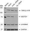
Knockdown of SESTD1 expression by specific SESTD1 siRNA. Western blot of HM1-C5 cells transfected with liposomes only (mock), 20 nm nonspecific control siRNA, or a 20 nm concentration of a specific SESTD1 siRNA pool (see “Experimental Procedures”). Blots were incubated with anti-SESTD1, anti-GFP, and anti-GAPDH antibodies. SESTD1-specific siRNA decreased SESTD1 protein by 85.5 ± 5.5% (n = 4, compared with mock-transfected cells) or 82.3 ± 5.3% (n = 4, compared with cells treated with nonspecific, non-silencing siRNA), whereas expression of TRPC5-YFP, an unrelated protein recognized by anti-SESTD1, as well as GAPDH expression were unchanged.
FIGURE 8.
FIGURE 8.
TRPC5 activity is reduced in HM1-C5 cells transfected with SESTD1 siRNA. Ratiometric measurements of [Ca2+]i in fura-2-loaded HM1-C5 cells transfected with 20 nm SESTD1 siRNA, nonspecific control siRNA, or liposomes only (Mock). Ca2+ release from internal stores was activated in Ca2+-free buffer by application of 10 μm carbachol (A) or 100 nm trypsin (B). Carbachol-induced (C) or trypsin-induced (D) TRPC5-mediated Ca2+ influx was assessed in standard Ca2+-containing buffer (2 mm). Shown are means ± S.E. of three independent experiments (each performed with n = 4–6 wells/experimental condition). Statistical analysis shows a selective reduction of agonist-induced TRPC5-mediated Ca2+ influx in SESTD1 siRNA-treated cells after termination of Ca2+ release (t = 121 s) as well as at the end of the experiment. (***, p < 0.001; *, p < 0.05 versus unselective siRNA; ***, p < 0.001 versus mock; analysis of variance).
FIGURE 9.
FIGURE 9.
Selective Ca2+-dependent binding of SESTD1 to phospholipids. PIP strips containing various immobilized phospholipids (left) were probed with GST-SESTD1 or GST in buffer containing 60 nm or 2.5 μm free Ca2+. Bound proteins were detected with anti-GST antibodies and visualized using horseradish peroxidase-conjugated secondary antibodies and enhanced chemiluminescence. LPA, lysophosphatidic acid; LPC, lysophosphatidylcholine; PI, phosphatidylinositol; PE, phosphatidylethanolamine; PC, phosphatidylcholine; S1P, sphingosine-1-phosphate; PS, phosphatidylserine; PA, phosphatidic acid.

Similar articles

Cited by

References

    1. Hofmann T., Schaefer M., Schultz G., Gudermann T. (2002) Proc. Natl. Acad. Sci. U.S.A. 99, 7461–7466 - PMC - PubMed
    1. Schaefer M., Plant T. D., Obukhov A. G., Hofmann T., Gudermann T., Schultz G. (2000) J. Biol. Chem. 275, 17517–17526 - PubMed
    1. Trebak M., Lemonnier L., DeHaven W. I., Wedel B. J., Bird G. S., Putney J. W., Jr. (2009) Pflugers Arch. 457, 757–769 - PMC - PubMed
    1. Flemming P. K., Dedman A. M., Xu S. Z., Li J., Zeng F., Naylor J., Benham C. D., Bateson A. N., Muraki K., Beech D. J. (2006) J. Biol. Chem. 281, 4977–4982 - PubMed
    1. Philipp S., Cavalié A., Freichel M., Wissenbach U., Zimmer S., Trost C., Marquart A., Murakami M., Flockerzi V. (1996) EMBO J. 15, 6166–6171 - PMC - PubMed

Publication types

MeSH terms