. 2010 Apr;23(4):279-88.
doi: 10.1093/protein/gzq003. Epub 2010 Feb 17.

Generating a panel of highly specific antibodies to 20 human SH2 domains by phage display

Affiliations

Generating a panel of highly specific antibodies to 20 human SH2 domains by phage display

K Pershad et al. Protein Eng Des Sel. 2010 Apr.

Abstract

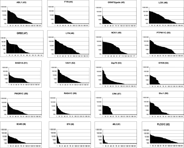
To demonstrate the utility of phage display in generating highly specific antibodies, affinity selections were conducted on 20 related Src Homology 2 (SH2) domains (ABL1, ABL2, BTK, BCAR3, CRK, FYN, GRB2, GRAP2, LYN, LCK, NCK1, PTPN11 C, PIK3R1 C, PLCgamma1 C, RASA1 C, SHC1, SH2D1A, SYK N, VAV1 and the tandem domains of ZAP70). The domains were expressed in Escherichia coli, purified and used in affinity selection experiments. In total, 1292/3800 of the resultant antibodies were shown to bind the target antigen. Of the 695 further evaluated in specificity ELISAs against all 20 SH2 domains, 379 antibodies were identified with unique specificity (i.e. monospecific). Sequence analysis revealed that there were at least 150 different clones with 1-19 different antibodies/antigen. This includes antibodies that distinguish between ABL1 and ABL2, despite their 89% sequence identity. Specificity was confirmed for many on protein arrays fabricated with 432 different proteins. Thus, even though the SH2 domains share a common three-dimensional structure and 20-89% identity at the primary structure level, we were able to isolate antibodies with exquisite specificity within this family of structurally related domains.


Figures

Fig. 1
Fig. 1
The family of SH2 domains. (A) Hypertree representation of all 120 human SH2 domains. A family tree of 120 SH2 domains in the human proteome is shown, with the 21 domains used in this study highlighted in red and underlined. The ZAP70 protein fragment used in this study contains two SH2 domains ZAP70_N and ZAP70_C, which are represented separately in the diagram. (B) The three-dimensional structure of ABL2. The structure of the SH2 domain of ABL2 is shown, with highlighting of residues differing between ABL1 and ABL2. The PyMOL Molecular Graphics System (DeLano Scientific, San Carlos, CA, USA) was used to generate a surface model of ABL2 from the PDB coordinates 2ECD (Kasai et al., doi:10.2210/pdb2ecd/pdb). (C) Sequence comparison of ABL1 and ABL2 SH2 domains. The ABL2 primary structure is shown indicating the 11 residues differing between ABL1 and ABL2.
Fig. 2
Fig. 2
SDS–PAGE analysis of the 20 purified human SH2 domains. (AC) The recombinant proteins were purified by IMAC and gel filtration. The proteins were resolved by SDS–PAGE gel to confirm their purity. Molecular weights of size standards are shown in kiloDaltons.
Fig. 3
Fig. 3
Primary screen of scFvs selected with each of the 20 SH2 domains. Affinity selections were carried out on 20 different human SH2 domain proteins and 190 antibodies, from the second round of selection, were tested for binding to each target. Binding of scFvs to the immobilized targets was quantified using europium-labeled anti-Flag secondary antibody. A graph is shown for each selection plotting the time-resolved fluorescence signal in intensity units (y-axis, logarithmic scale) for all 190 clones in that selection (x-axis). Antibodies are named according to their selection identification number which is shown in parentheses next to the target name (also used in Table I).
Fig. 4
Fig. 4
Binding specificity for the VAV1 binding scFvs against the other 19 SH2 domain proteins. (A) Twenty-one scFvs passing primary specificity screening were incubated with the specific target VAV1 and 19 other control SH2 domain proteins. The ELISA signal is represented as fluorescent intensity units on the y-axis (logarithmic scale) and the antibody name is shown on the x-axis. All the scFv clones were specific for VAV1 (specific signal >10) except clone 52_E12. scFv clones having identical sequences (as described in Table II), and indicated by letters a, b, and c. (B) Specificity plot showing specific ABL1 versus ABL2 clones. ELISA signal on ABL1 versus ABL2 is shown for clones selected on either antigen. In the case of ABL1 selections, 32 clones arose sharing a common heavy chain in combination with at least four different light chains (based on the CDR3 sequences of VH and VL). These are identified as groups a, b, c and clone 43_F12. Groups d, e and f represent other groupings of duplicate clones. Within Group a, three different isolates were identified with sequence differences outside of CDR3. Thus, Group a is further sub-divided into ai, aii and aiii. Group g represents the three clones selected on ABL2.
Fig. 5
Fig. 5
Specificity of antibody binding on protein microarrays. Microarrays spotted with 432 different proteins were probed with a range of antibodies. Examples show specific recognition of GRB2 (using 47_B02 at 6 µg/ml) SYK_N (using 054_A04 at 1 µg/ml) and RASA1_C (using 056_A10 at 1 µg/ml). In the case of NCK, the same antibody (067_F09) was used at either 13 or 1.2 µg/ml. Increased background was observed at the higher concentration. A green bar represents recognition of the correct antigen and a black bar shows cross-reactivity.

References

    1. Editorial. Nat. Methods. 2008;5:851.
    1. Haab B.B., Paulovich A.G., Anderson N.L., Clark A.M., Downing G.J., Hermjakob H., Labaer J., Uhlen M. Mol. Cell. Proteomics. 2006;5:1996–2007. - PubMed
    1. Huang H., et al. Mol. Cell. Proteomics. 2008;7:768–784. - PubMed
    1. Huber T., Steiner D., Rothlisberger D., Pluckthun A. J. Struct. Biol. 2007;159:206–221. - PubMed
    1. Kasai T., Koshiba S., Inoue M., Kigawa T., Yokoyama S. doi:10.2210/pdb2ecd/pdb. - DOI

Publication types

Substances