. 2010 May;84(9):4755-68.
doi: 10.1128/JVI.00851-09. Epub 2010 Feb 17.

Human T-lymphotropic virus type 1 transcription and chromatin-remodeling complexes

Affiliations

Human T-lymphotropic virus type 1 transcription and chromatin-remodeling complexes

Rebecca Easley et al. J Virol. 2010 May.

Abstract

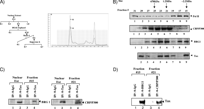
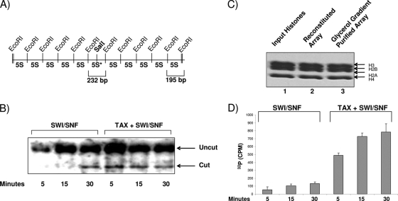
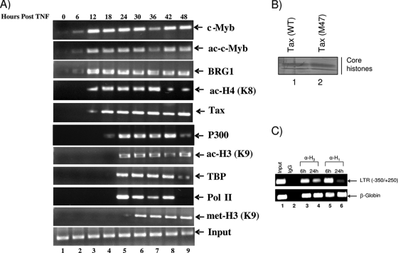
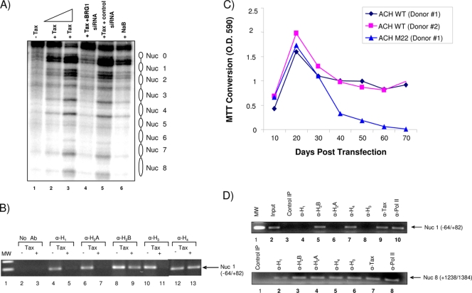
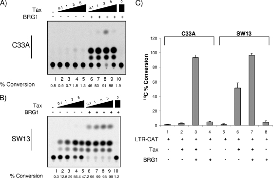
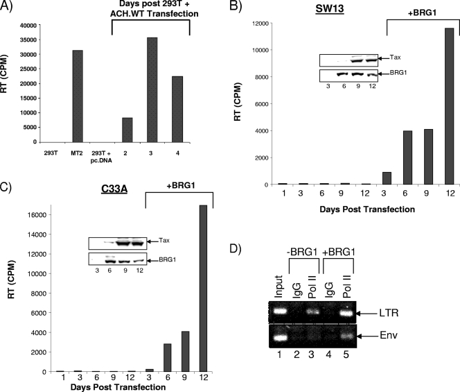
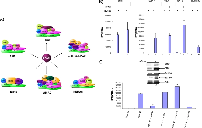
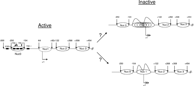
Human T-lymphotropic virus type 1 (HTLV-1) encodes the viral protein Tax, which is believed to act as a viral transactivator through its interactions with a variety of transcription factors, including CREB and NF-kappaB. As is the case for all retroviruses, the provirus is inserted into the host DNA, where nucleosomes are deposited to ensure efficient packaging. Nucleosomes act as roadblocks in transcription, making it difficult for RNA polymerase II (Pol II) to proceed toward the 3' end of the genome. Because of this, a variety of chromatin remodelers can act to modify nucleosomes, allowing for efficient transcription. While a number of covalent modifications are known to occur on histone tails in HTLV-1 infection (i.e., histone acetyltransferases [HATs], histone deacetylases [HDACs], and histone methyltransferases [HMTs]), evidence points to the use of chromatin remodelers that use energy from ATP hydrolysis to remodel nucleosomes. Here we confirm that BRG1, which is the core subunit of eight chromatin-remodeling complexes, is essential not only for Tax transactivation but also for viral replication. This is especially evident when wild-type infectious clones of HTLV-1 are used. BRG1 associates with Tax at the HTLV-1 long terminal repeat (LTR), and coexpression of BRG1 and Tax results in increased rates of transcription. The interaction of BRG1 with Tax additionally recruits the basal transcriptional machinery and removes some of the core histones from the nucleosome at the start site (Nuc 1). When using the BRG1-deficient cell lines SW13, C33A, and TSUPR1, we observed little viral transcription and no viral replication. Importantly, while these three cell lines do not express detectable levels of BRG1, much of the SWI/SNF complex remains assembled in the cells. Knockdown of BRG1 and associated SWI/SNF subunits suggests that the BRG1-utilizing SWI/SNF complex PBAF is responsible for HTLV-1 nucleosome remodeling. Finally, HTLV-1 infection of cell lines with a knockdown in BRG1 or the PBAF complex results in a significant reduction in viral production. Overall, we concluded that BRG1 is required for Tax transactivation and HTLV-1 viral production and that the PBAF complex appears to be responsible for nucleosome remodeling.


Figures

FIG. 1.
FIG. 1.
Association of Tax with CBP/p300 and SWI/SNF complexes. (A) (Left) Schematic representation of chromatographic steps using C8166 nuclear extracts. Nuclear extracts were passed through P11, DEAE Sephacel, Mono S, and Superose 6 sizing columns to obtain Tax and associated factors. (Right) Chromatogram of the fast protein liquid chromatography (FPLC) fractions, where peak A shows Superose 6 at 0.7 M salt and peak B shows 0.15 M salt. Fractions 13 to 17, which contain a high-molecular-weight complex, are fairly stable at either salt concentration. (B) Western blot analysis of Superose 6 column fractions using antibodies against Pol II, CBP/p300, Tax, and BRG1. Fractions were loaded and isolated at 0.7 M salt. Tax eluted at three distinct locations, including a small complex (120 kDa), a medium complex (1 MDa), and a large complex (∼2.2 MDa). (C) Immunoprecipitation using anti-Tax or control antibodies (IgG), followed by Western blot analysis for antibodies against BRG1 and CBP/p300. p300 is slightly larger than CBP (molecular weight, 265,000 [265K]) (68). A total of 330 μg of each fraction (fractions 15 and 21) was used for immunoprecipitation with 1.0 μg of the primary antibody. (D) Reciprocal IP of fraction 15 with anti-BRG1, as well as IP with anti-CBP/p300 from fraction 21, followed by Western blotting for Tax.
FIG. 2.
FIG. 2.
Functional interaction of Tax with the chromatin-remodeling complex in vitro. (A) Diagram of the 5S nucleosome positioning plasmid, bearing a unique SalI site close to the dyad axis of a reconstituted nucleosome in the middle of an array of 10 5S nucleosome positioning sequences. EcoRI sites are 195 bp apart, while the SalI 5S repeat is located on a 232-bp EcoRI fragment. Modified from reference . (B) Glycerol gradient-purified 5S chromatinized DNA was used in a SalI restriction enzyme accessibility assay using either SWI/SNF alone or Tax plus SWI/SNF. The reaction mixtures were incubated at 37°C. Reactions were stopped after 5, 15, and 30 min before samples were processed and run on an agarose gel. The DNA was run on a 1.2% gel and was then stained with ethidium bromide. The gels were then dried onto 3MM Whatman paper. The fraction of uncut DNA template on the array was obtained by phosphorimager analysis using ImageQuant software, by taking the ratio of uncut signal to the sum of cut and uncut signals present in the lane. (C) Presence of histones on DNA after gradient purification as shown by Coomassie blue staining. Glycerol gradient sedimentation of arrays was carried out on 10 to 30% linear gradients. Sedimentation velocity studies were performed on a Beckman XL-A analytical ultracentrifuge equipped with scanner options as described previously (43). Lane 1, input histones used for reconstitution arrays (2.5 μg); lanes 2 and 3, reconstituted DNA molecules before and after exposure to the glycerol gradient. (D) Similar to panel C, but the plasmid used contained an LTR luciferase construct, and following nucleosome assembly, samples were treated with SmaI and SacI and were run on an agarose gel (2.5%), followed by hybridization with a nick-translated probe spanning the area of the LTR from −52 to +1. Data represent the results of three experiments with the hybridized cut fragment and SWI/SNF or Tax/SWI/SNF.
FIG. 3.
FIG. 3.
HTLV-1 clone transfection and in vivo ChIP analysis. (A) Human 293T kidney fibroblasts were seeded to 60% confluence in 10-cm-diameter dishes and were transfected with ACH.WT (20 μg) or pBluescript KS (20 μg of empty vector) (data not shown). After transfection, the cells were maintained for 1 week, and the remaining live cells (latent cells) were treated with TNF to activate the virus. Samples were processed every 6 h following TNF treatment for ChIP analysis with appropriate antibodies. Cell culture supernatants were collected at various time points posttransfection, and virus particle production was monitored by a p19 enzyme-linked immunosorbent assay (ELISA) (data not shown). The input panel (positive-control PCR) shows the presence of HTLV-1 DNA in total chromosomal DNA prior to the ChIP assay. The primers for the ChIP assay spanned the region from −350 to +250 (U3/R/U5). The 24-h sample represents the start of activated transcription (recruitment of the nucleating factor TBP) by Tax. ac, acetylation; met, methylation. (B) Isolation of total histones from cells transfected with ACH.WT (20 μg) or an ACH.M47 (20 μg) mutant clone. Histones were isolated after 48 h and were run on a 4 to 20% gel, which was stained with Coomassie blue. (C) 293T cells were transfected with ACH.WT (20 μg) as for panel A. Samples were processed after either 6 or 24 h for ChIP analysis. Antibodies against histone H1 and H3 were used for ChIP, and recovered DNA was subsequently used for PCR with primers against the LTR (from −350 to +250) or the beta-globin promoter.
FIG. 4.
FIG. 4.
Determination of translational positioning of HTLV-1 nucleosomes and absence of various histones in transfected H1GFP cells and PBMCs. (A) DNase I treatment of permeabilized H1GFP (HTLV-1 LTR-GFP) cells for mapping of DNase I-hypersensitive sites. Cells were grown to 70% confluence and were then either left untransfected (lane 1); transfected with increasing amounts of Tax alone (lane 2, 5 μg; lane 3, 10 μg), five wild-type BRG1-specific siRNAs (20 μg) plus Tax (10 μg) (lane 4), or five mutant BRG1 siRNAs (20 μg) plus Tax (10 μg) (lane 5); or treated with NaB (10 μM). Forty-eight hours later, cells were prepared for a DNase I hypersensitivity assay, size fractionated on an agarose gel, and transferred to a membrane. The nick-translated H1GFP fragment (5 μg) was used as a hybridization probe for Southern blotting. (B) Effect of Tax on histone eviction after Tax treatment. H1GFP cells were either left untreated or transfected with a Tax plasmid using Lipofectamine. Forty-eight hours later, samples were processed for ChIP using various antibodies against histones. Primers for the +1 start site (Nuc 1 area) spanned the area from −64 to +82. (C) Human PBMCs from two healthy donors were first purified by Ficoll-Paque centrifugation and then activated with PHA (10 μg/ml) and recombinant human IL-2 (50 U/ml). The cells were transfected with double-CsCl-purified ACH.WT (40 μg) using the Amaxa reagent. The cells were then cultured in cRPMI supplemented with PHA (5 μg/ml) and IL-2 for ∼6 weeks, after which they were cultured in the same medium without PHA. At various time points, cell culture supernatants were collected for p19 determination (data not shown), and relative cellular viability was assayed by MTT conversion assays on 100-μl aliquots of cells. Cells that survived for 70 days were ∼90% infected with HTLV-1. O.D. 590, optical density at 590 nm. (D) ChIP assays with samples from donor 1 PBMCs transfected with the wild-type clone ACH.WT. ChIPs were performed with 70-day samples using various antibodies. PCR amplification was performed for both the Nuc 1 (+1 area) and Nuc 8 (Gag) regions.
FIG. 5.
FIG. 5.
Effect of BRG1 on HTLV-1 LTR Tax-activated transcription in mutant cells. (A and B) Titration of Tax (0.1, 1, 3, and 5 μg) either alone (lanes 2 to 5) or with a BRG1 plasmid (5 μg) (lanes 6 to 9). A total of 5 × 10⁶ C33A (A) or SW13 (B) cells were transfected for 3 days and were subsequently processed for CAT assays (50 μg of total extract). Lanes 1 serve as a negative control (HTLV-1-LTR-CAT and no Tax), and lanes 10 also serve as a control for BRG1 (5 μg of a BRG1 dominant-negative mutant [38]). BRG1 enhanced the level of activation dramatically even with minimal concentrations of Tax (i.e., lanes 6 in both panels). (C) LTR-CAT was transfected either with BRG1 alone or with BRG1 plus Tax (3 μg) into either C33A or SW13 cells, as for panels A and B. The data represent three independent replicates. %Conversion, %conversion from unacetylated to mono- or diacetylated chloramphenicol.
FIG. 6.
FIG. 6.
Lack of HTLV-1 viral replication in BRG1 mutant cells. (A) 293T cells, used as a control, were transfected with the HTLV-1 wild-type clone, ACH.WT. Supernatants from cells infected with HTLV-1 MT2 (20 μl) and treated with TNF or from 293T cells transfected with an empty vector (supernatants after 4 days) served as positive and negative controls, respectively. Optimal HTLV-1 replication occurred on day 3. (B and C) Both SW13 and C33A BRG1 mutant cells were transfected with either the pcDNA3 vector (data not shown) or the ACH.WT plasmid (20 μg), and experiments were carried out to day 12. Day 1 is a negative control with no transfection. Viral RT was present only in the supernatants of BRG1-transfected cells and peaked at day 12 in either cell type. All experiments in panels B and C were performed in triplicate. Western blotting for BRG1 and actin of samples from SW13 or C33A cells transfected with a mock plasmid (left lanes 1, 3, 9, and 12) or a BRG1 plasmid on days 3, 6, 9, and 12 (right lanes and insets). (D) C33A cells from panel C at day 12 were processed for ChIP experiments using either an IgG control or the anti-Pol II antibody 8WG16. The LTR primers spanned the region from −350 to +250 (U3/R/U5), and the Env primers spanned the region from +8990 to +9450.
FIG. 7.
FIG. 7.
Effects of various siRNAs against SWI/SNF components on the wild-type HTLV-1 LTR. (A) BRG1-associated complexes. Brahma-related gene-1 (BRG1) is a catalytic subunit found in a variety of chromatin-remodeling complexes, including the SWI/SNF complexes BAF and PBAF, as well as WINAC, NCoR, mSin3A/HDAC, and NUMAC. The various complexes share other subunits that have a variety of activities, color coded as follows: green, core components; pink, nuclear receptor association; gold, DNA replication; light green, actin-related complexes; blue, Ini1/Baf47/SNF5; purple, other functions (63). As chromatin remodelers, these complexes can act either as transcriptional activators, as transcriptional repressors, or as both. For example, NUMAC acts as a transcriptional activator, while the mSin3A/HDAC complex acts as a transcriptional repressor. SWI/SNF complexes can act as either repressors or activators. (B) RT assay of supernatants (10 μl) from 293T cells (positive control), BRG1 mutant (TSUPR1, C33A, and SW13) cells, and Baf180 mutant (HCC1143) cells. Cells were transfected with the ACH.WT plasmid (20 μg) and were grown for 6 days. Supernatants were collected at day 6 and were then processed for the RT assay. BRG1 (10 μg), along with ACH.WT, was also transfected into five cell types (lanes 2, 4, 6, 8, and 10). (C) 293T cells were transfected with various siRNAs, including siBRG1, siBRM, siBaf250, and siBaf180 (300 nM), along with ACH.WT (20 μg). Samples were processed for RT at day 6. (Inset) Cell pellets were processed for Western blotting with antibodies against BRG1 and various other components. The actin panel represents cells transfected with siBRG1. Data represent one replicate of three independent experiments.
FIG. 8.
FIG. 8.
Model of nucleosomal arrays on the Tax-activated HTLV-1 LTR in vivo. (Left) In the presence of Tax, there are well-defined translationally positioned nucleosomal arrays on the active promoter, as seen by the presence of DNase I-hypersensitive sites and the removal of histones H1, H2A, and H3 at the transcriptional start site region. (Right) DNase I cleavage analysis indicates that the inactive promoter does not contain translationally positioned nucleosomal arrays. This is indicated by the lack of DNase I-hypersensitive sites and the presence of all core histones as well as H1. Currently it is not clear whether the Nuc 1 area at the transcriptional start site contains multiple arrays that are not phased (top right) or whether there is only one nucleosome that makes the DNA inaccessible for transcription factor and/or Pol II occupancy (bottom right). The arrows at both ends of each nucleosome represent the beginning of the hypersensitive site. The removal of some of the histones is shown as a dotted oval around Nuc1.

References

    1. Agbottah, E., L. Deng, L. O. Dannenberg, A. Pumfery, and F. Kashanchi. 2006. Effect of SWI/SNF chromatin remodeling complex on HIV-1 Tat activated transcription. Retrovirology 3:48. - PMC - PubMed
    1. Akagi, T., H. Ono, and K. Shimotohno. 1996. Expression of cell-cycle regulatory genes in HTLV-I infected T-cell lines: possible involvement of Tax1 in the altered expression of cyclin D2, p18Ink4 and p21Waf1/Cip1/Sdi1. Oncogene 12:1645-1652. - PubMed
    1. Bannister, A. J., and T. Kouzarides. 1996. The CBP co-activator is a histone acetyltransferase. Nature 384:641-643. - PubMed
    1. Brower-Toland, B., D. A. Wacker, R. M. Fulbright, J. T. Lis, W. L. Kraus, and M. D. Wang. 2005. Specific contributions of histone tails and their acetylation to the mechanical stability of nucleosomes. J. Mol. Biol. 346:135-146. - PubMed
    1. Caron, C., R. Rousset, C. Beraud, V. Moncollin, J. M. Egly, and P. Jalinot. 1993. Functional and biochemical interaction of the HTLV-I Tax1 transactivator with TBP. EMBO J. 12:4269-4278. - PMC - PubMed

Publication types