. 2010 May;84(9):4682-96.
doi: 10.1128/JVI.00126-10. Epub 2010 Feb 17.

Recruitment of herpes simplex virus type 1 immediate-early protein ICP0 to the virus particle

Affiliations

Recruitment of herpes simplex virus type 1 immediate-early protein ICP0 to the virus particle

Kevin Maringer et al. J Virol. 2010 May.

Abstract

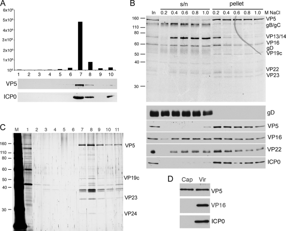
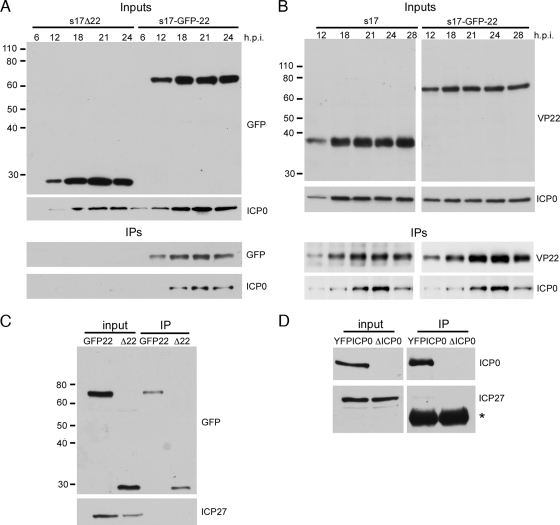
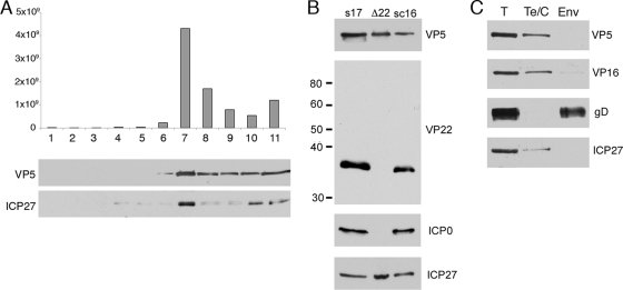
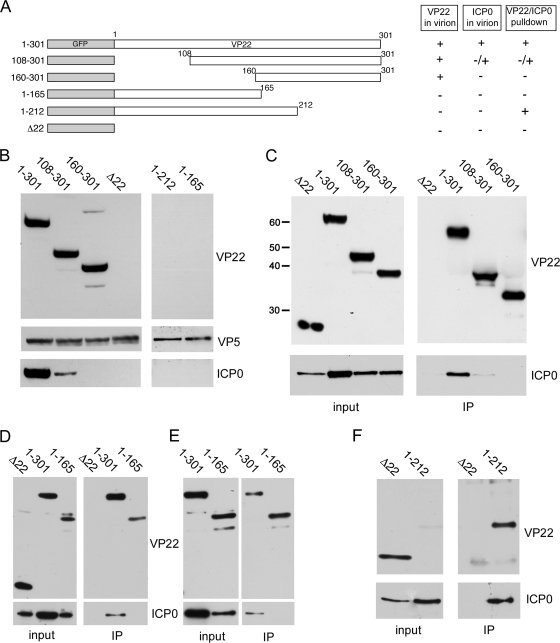
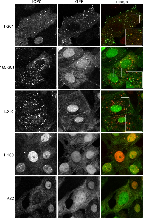
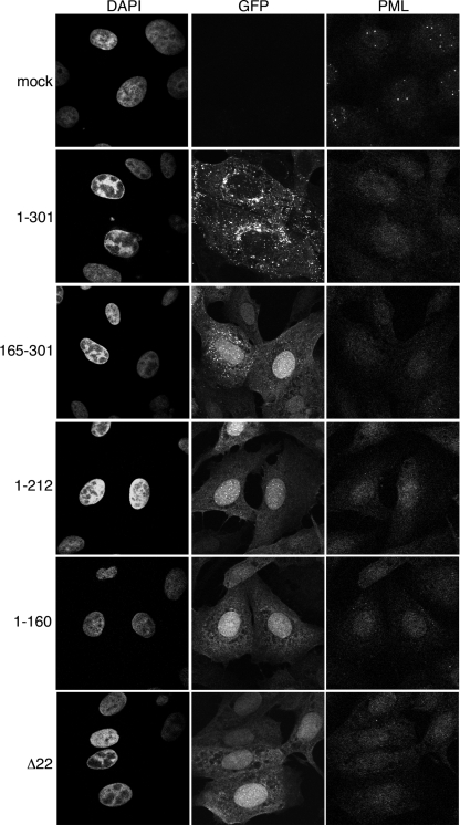
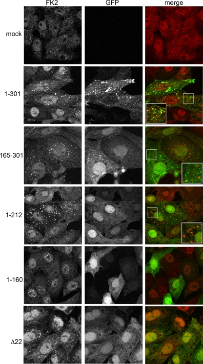
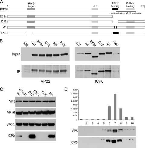
Although the herpes simplex virus type 1 (HSV-1) tegument is comprised of a large number of viral and cellular proteins, how and where in the cell these proteins are recruited into the virus structure is poorly understood. We have shown previously that the immediate-early gene product ICP0 is packaged by a mechanism dependent on the major tegument protein VP22, while others have shown a requirement for ICP27. We now extend our studies to show that ICP0 packaging correlates directly with the ability of ICP0 to complex with VP22 in infected cells. ICP27 is not, however, present in this VP22-ICP0 complex but is packaged into the virion in a VP22- and ICP0-independent manner. Biochemical fractionation of virions indicated that ICP0 associates tightly with the virus capsid, but intranuclear capsids contained no detectable ICP0. The RING finger domain of ICP0 and the N terminus of VP22 were both shown to be essential but not sufficient for ICP0 packaging and complex formation. Strikingly, however, the N-terminal region of VP22, while unable to form a complex with ICP0, inhibited its translocation from the nucleus to the cytoplasm. PML degradation by ICP0 was efficient in cells infected with this VP22 mutant virus, confirming that ICP0 retains activity. Hence, we would suggest that VP22 is an important molecular partner of ICP0 that controls at least one of its activities: its assembly into the virion. Moreover, we propose that the pathway by which VP22 recruits ICP0 to the virion may begin in the nucleus prior to ICP0 translocation to its final site of assembly in the cytoplasm.


Figures

FIG. 1.
FIG. 1.
ICP0 is an HSV-1 tegument protein. (A) Extracellular s17 virions were centrifuged through a 5 to 15% Ficoll gradient, and fractions were taken across the gradient. Each sample was analyzed for infectivity (top) or subjected to Western blotting for the major capsid protein VP5 or ICP0. (B) Purified extracellular s17 virions were fractionated using detergent and increasing concentrations of NaCl. Soluble (s/n) and insoluble (pellet) fractions were separated by pelleting and then analyzed by SDS-PAGE followed by Coomassie blue staining (top panel) or Western blotting for a range of virus structural proteins. The sizes of the molecular weight markers (in thousands) are indicated on the left. (C) Intranuclear capsids from s17-infected cells were centrifuged through a 15 to 50% sucrose gradient, and fractions were collected across the gradient. Each sample was analyzed by SDS-PAGE followed by silver staining. (D) Equivalent amounts of intact s17 virions and s17 capsids from fraction 8, based on their VP5 content, were analyzed by Western blotting for VP5, VP16, and ICP0.
FIG. 2.
FIG. 2.
VP22 and ICP0 form a complex in infected cells. (A) Vero cells infected with s17 expressing GFP in place of VP22 (s17Δ22) or expressing GFP-tagged VP22 (s17-GFP-22) at a multiplicity of infection of 1 were harvested at times ranging between 6 and 24 h after infection, and immunoprecipitations carried out with a VP22 polyclonal antibody. The original input samples and resulting complexes were analyzed by Western blotting for GFP or ICP0. The sizes of the molecular weight markers (in thousands) are indicated on the left. (B) As described in the legend for panel A, but using s17 and s17-GFP-22 viruses. (C) Vero cells infected with the same viruses as those shown in panel A were harvested 20 h after infection and immunoprecipitated with a polyclonal anti-GFP antibody. The resulting complexes were analyzed by Western blotting for GFP and ICP27. (D) Vero cells infected with either s17 expressing YFP-tagged ICP0 or the ICP0 deletion virus dl1403 (ΔICP0) were treated as described in the legend for panel C and analyzed by Western blotting for ICP0 and ICP27. Asterisk denotes polyclonal heavy chain.
FIG. 3.
FIG. 3.
ICP27 is packaged into the HSV-1 tegument in a VP22-independent mechanism. (A) Extracellular HFEM virions were centrifuged through a 5 to 15% Ficoll gradient, and fractions taken across the gradient. Each sample was analyzed for infectivity (top) or subjected to Western blotting for the major capsid protein VP5 or ICP27. (B) Approximately equivalent amounts of s17, s17Δ22, and sc16 virions were analyzed by Western blotting for VP5, VP22, ICP0 and ICP27. The sizes of the molecular weight markers (in thousands) are indicated on the left. (C) Strain HFEM virions were fractionated into envelope (Env) and tegument-capsid (Te/C) fractions using detergent and 0.1 M NaCl. Soluble and insoluble fractions were separated by pelleting and then analyzed by Western blotting for the capsid protein VP5, the tegument protein VP16, the envelope protein gD, and ICP27.
FIG. 4.
FIG. 4.
The N terminus of VP22 is required for ICP0 assembly into the virion. (A) Line drawing of the VP22 truncation mutants used in this study. (B) Equivalent amounts of virions from the viruses described in the legend for panel A were analyzed by Western blotting for VP22, VP5, and ICP0. (C) Immunoprecipitations using a polyclonal GFP antibody were carried out on Vero cells infected with HSV-1 expressing full-length GFP-22 (1 to 301), the two N-terminal VP22 truncation mutants (108 to 301 and 160 to 301), or GFP in place of VP22 (Δ22), 20 h after infection at a multiplicity of infection of 1. The resulting complexes were analyzed by Western blotting for the C terminus of VP22 or ICP0. The sizes of the molecular weight markers (in thousands) are indicated on the left. (D and E) As described in the legend for panel C, using cells infected with HSV-1 expressing full-length VP22 (1 to 301), the N-terminal half of VP22 (1 to 165), or GFP in place of VP22 (Δ22). Western blots were carried out using antibodies for the N terminus of VP22 or ICP0. (F) As described in the legend for panel D, using cells infected with HSV-1 expressing GFP in place of VP22 (Δ22) or the C-terminal truncation (1 to 212).
FIG. 5.
FIG. 5.
ICP0 localizes to cytoplasmic domains in the absence of interacting VP22. Vero cells on coverslips were infected with the GFP-expressing viruses described in the legend for Fig. 4A at a multiplicity of infection of 10 and fixed with 4% paraformaldehyde 6 h later. Immunofluorescence was carried out for ICP0 (red).
FIG. 6.
FIG. 6.
PML is dispersed from ND10s in all VP22 mutant virus infections. Vero cells on coverslips were either uninfected (mock) or infected with the GFP-expressing viruses described in the legend for Fig. 4A at a multiplicity of infection of 10 and fixed with 4% paraformaldehyde 6 h later. Immunofluorescence was carried out for PML, and nuclei were stained with DAPI.
FIG. 7.
FIG. 7.
ICP0 induces colocalizing, conjugated ubiquitin in all VP22 mutant virus infections. Vero cells on coverslips were either uninfected (mock) or infected with the GFP-expressing viruses described in the legend for Fig. 4A at a multiplicity of infection of 10 and fixed with 4% paraformaldehyde 6 h later. Immunofluorescence was carried out for conjugated ubiquitin using antibody FK2 (red).
FIG. 8.
FIG. 8.
Virion assembly of ICP0 requires its RING finger and its C terminus. (A) Line drawing of the ICP0 mutants used in this study. (B) U2OS cells were infected with all the viruses shown in panel A together with the Δ22 mutant virus at a multiplicity of infection of 1 and harvested 20 h later. Immunoprecipitations were carried out using a VP22 antibody, and the resulting complexes were analyzed by Western blotting for VP22 and ICP0. (C) Equivalent amounts of extracellular virions from the viruses shown in panel A, together with the ICP0 deletion mutant (dl1403), were analyzed by Western blotting for VP5, VP16, VP22, and ICP0. (D) Extracellular virions from the USP7 binding site mutant D12 were centrifuged through a 5 to 15% Ficoll gradient, and fractions were taken across the gradient. Each sample was analyzed for infectivity (top) or subjected to Western blotting for the major capsid protein VP5 or ICP0.

Similar articles

Cited by

References

    1. Ackermann, M., D. K. Braun, L. Pereira, and B. Roizman. 1984. Characterization of herpes simplex virus 1 alpha proteins 0, 4, and 27 with monoclonal antibodies. J. Virol. 52:108-118. - PMC - PubMed
    1. Batterson, W., and B. Roizman. 1983. Characterization of the herpes simplex virion-associated factor responsible for the induction of alpha genes. J. Virol. 46:371-377. - PMC - PubMed
    1. Boutell, C., M. Canning, A. Orr, and R. D. Everett. 2005. Reciprocal activities between herpes simplex virus type 1 regulatory protein ICP0, a ubiquitin E3 ligase, and ubiquitin-specific protease USP7. J. Virol. 79:12342-12354. - PMC - PubMed
    1. Boutell, C., A. Orr, and R. D. Everett. 2003. PML residue lysine 160 is required for the degradation of PML induced by herpes simplex virus type 1 regulatory protein ICP0. J. Virol. 77:8686-8694. - PMC - PubMed
    1. Boutell, C., S. Sadis, and R. D. Everett. 2002. Herpes simplex virus type 1 immediate-early protein ICP0 and its isolated RING finger domain act as ubiquitin E3 ligases in vitro. J. Virol. 76:841-850. - PMC - PubMed

Publication types

MeSH terms

Substances

LinkOut - more resources