

Role of herpes simplex virus ICP0 in the transactivation of genes introduced by infection or transfection: a reappraisal
- PMID: 20164233
- PMCID: PMC2863771
- DOI: 10.1128/JVI.02585-09
Role of herpes simplex virus ICP0 in the transactivation of genes introduced by infection or transfection: a reappraisal
Abstract
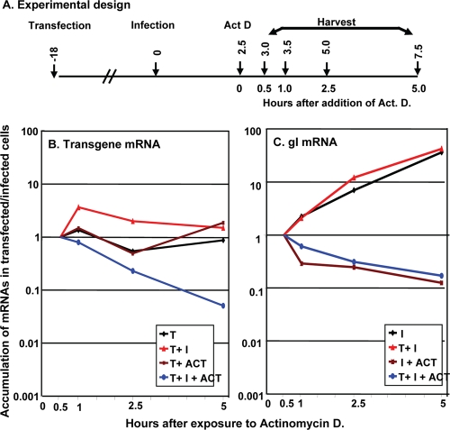
ICP0, a promiscuous transactivator that enhances the expression of genes introduced by infection or transfection, functions in both nucleus and cytoplasm. The nuclear functions include degradation and dispersal of ND10 bodies and suppression of silencing of viral DNA. Subsequently, ICP0 shifts to the cytoplasm. Transfection of DNA prior to infection has no effect on the localization of ICP0 in cells that are efficient expressers of transgenes (e.g., Vero and HEK293) but results in delayed cytoplasmic localization of ICP0 in cells (e.g., HEp-2 and HEL) that are poor transgene expressers. Here, we examined by real-time PCR (qPCR) the accumulation of a transgene and of viral gI mRNAs in Vero or HEp-2 cells that were transfected and then infected with wild-type or DeltaICP0 mutant viruses. The accumulation of transgene mRNA was unaffected by a DeltaICP0 mutant, gradually increased in HEp-2 cells, but increased and then decreased in Vero cells infected with wild-type virus. In both cell lines, accumulation of gI mRNA increased with time and was less affected by the transfected DNA in Vero cells than in HEp-2 cells. The relative kinetics of mRNA accumulation reflected continued synthesis and degradation of the transgene and gI mRNAs. We conclude that the role of ICP0 is to render the DNA templates introduced by transfection or infection accessible by transcriptional factors, that the two cell lines differ with respect to the transcription-ready status of entering foreign DNA in the nucleus, and that ICP0 is not per se the recruiter of transcriptional factors to the accessible DNA templates.
Figures


Similar articles
-
Activation of gene expression by herpes simplex virus type 1 ICP0 occurs at the level of mRNA synthesis.J Virol. 1997 Sep;71(9):6850-62. doi: 10.1128/JVI.71.9.6850-6862.1997. J Virol. 1997. PMID: 9261410 Free PMC article.
-
Overexpression of the herpes simplex virus type 1 immediate-early regulatory protein, ICP27, is responsible for the aberrant localization of ICP0 and mutant forms of ICP4 in ICP4 mutant virus-infected cells.J Virol. 1996 Aug;70(8):5346-56. doi: 10.1128/JVI.70.8.5346-5356.1996. J Virol. 1996. PMID: 8764045 Free PMC article.
-
Herpes simplex virus-infected cell protein 0 blocks the silencing of viral DNA by dissociating histone deacetylases from the CoREST-REST complex.Proc Natl Acad Sci U S A. 2007 Oct 23;104(43):17134-9. doi: 10.1073/pnas.0707266104. Epub 2007 Oct 15. Proc Natl Acad Sci U S A. 2007. PMID: 17939992 Free PMC article.
-
Herpes simplex virus type 1 ICP0 plays a critical role in the de novo synthesis of infectious virus following transfection of viral DNA.J Virol. 1989 Nov;63(11):4579-89. doi: 10.1128/JVI.63.11.4579-4589.1989. J Virol. 1989. PMID: 2552142 Free PMC article.
-
ICP0 enables and monitors the function of D cyclins in herpes simplex virus 1 infected cells.Proc Natl Acad Sci U S A. 2009 Aug 25;106(34):14576-80. doi: 10.1073/pnas.0906905106. Epub 2009 Aug 12. Proc Natl Acad Sci U S A. 2009. PMID: 19706544 Free PMC article.
Cited by
-
Neuron-associated retroelement-derived protein Arc/Arg3.1 assists in the early stages of alphaherpesvirus infection in human neuronal cells.PLoS One. 2024 Dec 12;19(12):e0314980. doi: 10.1371/journal.pone.0314980. eCollection 2024. PLoS One. 2024. PMID: 39666775 Free PMC article.
-
A Revision of Herpes Simplex Virus Type 1 Transcription: First, Repress; Then, Express.Microorganisms. 2024 Jan 26;12(2):262. doi: 10.3390/microorganisms12020262. Microorganisms. 2024. PMID: 38399666 Free PMC article. Review.
-
Use of biotinylated plasmid DNA as a surrogate for HSV DNA to identify proteins that repress or activate viral gene expression.Proc Natl Acad Sci U S A. 2012 Dec 18;109(51):E3549-57. doi: 10.1073/pnas.1218783109. Epub 2012 Dec 5. Proc Natl Acad Sci U S A. 2012. PMID: 23223531 Free PMC article.
-
The ICP0 Protein of Herpes Simplex Virus 1 (HSV-1) Downregulates Major Autophagy Adaptor Proteins Sequestosome 1 and Optineurin during the Early Stages of HSV-1 Infection.J Virol. 2019 Oct 15;93(21):e01258-19. doi: 10.1128/JVI.01258-19. Print 2019 Nov 1. J Virol. 2019. PMID: 31375597 Free PMC article.
-
Interwoven roles of cyclin D3 and cdk4 recruited by ICP0 and ICP4 in the expression of herpes simplex virus genes.J Virol. 2010 Oct;84(19):9709-17. doi: 10.1128/JVI.01050-10. Epub 2010 Jul 21. J Virol. 2010. PMID: 20660182 Free PMC article.
References
Publication types
MeSH terms
Substances
Grants and funding
LinkOut - more resources
Full Text Sources
