. 2010 May;84(9):4124-35.
doi: 10.1128/JVI.02319-09. Epub 2010 Feb 17.

Herpes simplex virus 1 regulatory protein ICP27 undergoes a head-to-tail intramolecular interaction

Affiliations

Herpes simplex virus 1 regulatory protein ICP27 undergoes a head-to-tail intramolecular interaction

Felicia P Hernandez et al. J Virol. 2010 May.

Abstract

Herpes simplex virus type 1 (HSV-1) regulatory protein ICP27 is a multifunction functional protein that interacts with many cellular proteins. A number of the proteins with which ICP27 interacts require that both the N and C termini of ICP27 are intact. These include RNA polymerase II, TAP/NXF1, and Hsc70. We tested the possibility that the N and C termini of ICP27 could undergo a head-to-tail intramolecular interaction that exists in open and closed configurations for different binding partners. Here, we show by bimolecular fluorescence complementation (BiFC) assays and fluorescence resonance energy transfer (FRET) by acceptor photobleaching that ICP27 undergoes a head-to-tail intramolecular interaction but not head-to-tail or tail-to-tail intermolecular interactions. Substitution mutations in the N or C termini showed that the leucine-rich region (LRR) in the N terminus and the zinc finger-like region in the C terminus must be intact for intramolecular interactions. A recombinant virus, vNC-Venus-ICP27, was constructed, and this virus was severely impaired for virus replication. The expression of NC-Venus-ICP27 protein was delayed compared to ICP27 expression in wild-type HSV-1 infection, but NC-Venus-ICP27 was abundantly expressed at late times of infection. Because the renaturation of the Venus fluorescent protein results in a covalent bonding of the two halves of the Venus molecule, the head-to-tail interaction of NC-Venus-ICP27 locks ICP27 in a closed configuration. We suggest that the population of locked ICP27 molecules is not able to undergo further protein-protein interactions.


Figures

FIG. 1.
FIG. 1.
BiFC analysis shows that ICP27 undergoes a head-to-tail intramolecular interaction. RSF cells were transfected with plasmid DNA from ICP27-Venus fusion constructs N-Venus/ICP27/C-Venus, N-Venus/ICP27 (L8A, L11A, L15A)/C-Venus, N-Venus/ICP27 (C483S, C488S)/C-Venus, and N-Venus/ICP27 (S16A, S18A) or were cotransfected with N-Venus-ICP27 and ICP27-C-Venus or with ICP27-N-Venus and ICP27-C-Venus as indicated. Eighteen hours after transfection, cells were infected with 27-LacZ at an MOI of 10. Venus fluorescence was visualized directly at 4 and 8 h after infection.
FIG. 2.
FIG. 2.
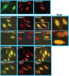
Immunofluorescence analysis indicates that the ICP27-Venus fusion constructs are expressed and localized to the nucleus. RSF cells were transfected with plasmid DNA from a wild-type ICP27-containing plasmid and from the ICP27-Venus fusion constructs indicated. Eighteen hours after transfection, cells were infected with 27-LacZ at an MOI of 10 for 4 and 8 h as indicated. Wild-type ICP27 was stained with anti-ICP27 antibody P1119, as was ICP27-C-Venus and ICP27-N-Venus. N-Venus-ICP27, N-Venus/ICP27 (C483S, C488S)/C-Venus, and N-Venus/ICP27 (L8A, L11A, L15A)/C-Venus were stained with an anti-GFP antibody that recognizes an epitope also present in Venus. The P1119 anti-ICP27 antibody recognizes an epitope at the N terminus of ICP27 (23) that is masked by the N-Venus fusion protein. Venus yellow fluorescence was visualized directly for N-Venus/ICP27/C-Venus.
FIG. 3.
FIG. 3.
ICP27-Venus fusion proteins are expressed to similar levels in transfected cells. RSF cells were transfected with the ICP27-Venus fusion constructs as indicated or with pSG130B/S, which expresses wild-type ICP27. Twenty-four hours later, cells were infected with 27-LacZ for 8 h, at which time samples were harvested. Western blot analysis was performed with anti-ICP27, anti-GFP, and anti-β-actin antibodies. The green arrow in the left panel points to the NC-Venus-ICP27 fusion proteins, and the red arrow points to wild-type ICP27. The positions of N-Venus/ICP27 and ICP27/C-Venus are indicated in the right panel. N-Venus/ICP27 migrates more slowly than ICP27/C-Venus because of the protein linker that was used in the construction of the N-Venus fusion protein. The asterisk in the left panel denotes a band that is likely a degradation product of the Venus-ICP27 fusion proteins and which migrated slightly faster than actin.
FIG. 4.
FIG. 4.
ICP27 intramolecular interaction is not competed for by wild-type ICP27. RSF cells were transfected with N-Venus/ICP27/C-Venus or N-YFP/ICP27/C-YFP or were cotransfected with N-Venus-ICP27 and ICP27-C-Venus as indicated. Cells were infected 18 h later with HSV-1 KOS or 27-LacZ at an MOI of 10 for 4 and 8 h as indicated. Venus fluorescence was visualized directly.
FIG. 5.
FIG. 5.
FRET by acceptor photobleaching demonstrates that ICP27 undergoes a head-to-tail intramolecular interaction but not an intermolecular interaction. (A and B) RSF cells were transfected with N-CFP/ICP27/C-YFP DNA. Cells were infected 24 h after transfection with 27-LacZ at an MOI of 10. Cells were fixed at 8 h after infection. (C) RSF cells were transfected with N-CFP/ICP27 and ICP27/C-YFP DNA. Cells were infected 24 h after transfection with 27-LacZ at an MOI of 10. Cells were fixed at 8 h after infection. CFP-YFP FRET by acceptor photobleaching was performed using an LSM 510 Meta confocal microscope as described by Karpova et al. (18) and detailed in Materials and Methods. The white circles indicate the specific area within each cell that was bleached at 514 nm. FRET efficiency, which is plotted to the right of the fluorescent images, was calculated as follows: FRET (%) = 1 − (donor before bleach − background before bleach)/(donor after bleach − background after bleach) × 100.
FIG. 6.
FIG. 6.
Recombinant virus v-NC-Venus-ICP27 is defective in viral replication. The v-NC-Venus-ICP27 virus was constructed by the marker transfer of N-Venus/ICP27/C-Venus into 27-LacZ (40), replacing the 27-LacZ locus. (A) One-step growth curves were performed by infecting Vero cells with KOS, 27-GFP, and vNC-Venus-ICP27 at an MOI of 1. The experiments were performed in triplicate, and viral titers at 4, 8, 16, and 24 h were determined by plaque assays on Vero cells. (B) Vero cells were infected with v-NC-Venus-ICP27 and v-N-YFP-ICP27 at an MOI of 10. Venus and YFP fluorescence were visualized directly on living cells at 4, 6, and 8 h after infection. (C) Vero cells were infected with KOS, v-N-YFP-ICP27, and v-NC-Venus-ICP27 at an MOI of 10. Western blot analysis was performed on protein samples isolated at 4, 8, and 12 h after infection. The blot was probed with anti-ICP27 and anti-GFP antibodies and with β-actin antibody as a loading control. The red arrows indicate the position of wild-type ICP27. The green arrows indicate the position of N-YFP-ICP27 and N-Venus/ICP27/C-Venus. The asterisks denote ICP27 protein degradation products in the N-YFP-ICP27 lanes. The position of molecular size markers is shown to the left of the gel. (D) Vero cells infected with KOS, vN-YFP-ICP27, and v-NV-Venus-ICP27 at an MOI of 10 were harvested at 4, 8, and 12 h after infection, and Western blot analysis was performed as described previously (42). The blots were probed with antibodies directed to ICP4, gC, gD, and β-actin as a loading control.
FIG. 7.
FIG. 7.
Replication compartment formation is hindered during infection with v-NC-Venus-ICP27. RSF cells were infected with KOS, vN-YFP-ICP27, and v-NC-Venus-ICP27 at an MOI of 10 for 8 h, at which time cells were fixed. Immunofluorescent staining was performed with anti-ICP27 antibody for KOS-infected cells, anti-RNAP II antibody ARNA3, and anti-ICP4 antibody. YFP and Venus fluorescence was visualized directly. White rectangles indicate the region of the merged images that is shown in the zoomed images to the right. White arrows point to replication compartments, and yellow arrows point to small prereplication sites.
FIG. 8.
FIG. 8.
Overexpression of TAP/NXF1 results in the export of NC-Venus-ICP27 to the cytoplasm. RSF cells either were not transfected or were transfected with CFP-TAP DNA as indicated. Twenty-four hours later, cells were infected with KOS, vN-YFP-ICP27, and v-NC-Venus-ICP27 at an MOI of 10. Cells were fixed at 8 and 12 h after infection. KOS-infected cells were stained with anti-ICP27 antibody, and CFP, YFP, and Venus fluorescence were visualized directly.

Similar articles

Cited by

References

    1. Bachi, A., I. C. Braun, J. P. Rodrigues, N. Pante, K. Ribbeck, C. von Kobbe, U. Kutay, M. Wilm, D. Gorlich, M. Carmo-Fonseca, and E. Izaurralde. 2000. The C-terminal domain of TAP interacts with the nuclear pore complex and promotes export of specific CTE-bearing RNA substrates. RNA 6:136-158. - PMC - PubMed
    1. Bryant, H. E., S. Wadd, A. I. Lamond, S. J. Silverstein, and J. B. Clements. 2001. Herpes simplex virus IE63 (ICP27) protein interacts with spliceosome-associated protein 145 and inhibits splicing prior to the first catalytic step. J. Virol. 75:4376-4385. - PMC - PubMed
    1. Burch, A. D., and S. K. Weller. 2004. Nuclear sequestration of cellular chaperone and proteasomal machinery during herpes simplex virus type 1 infection. J. Virol. 78:7175-7185. - PMC - PubMed
    1. Chattopadhyay, S., and S. K. Weller. 2007. Direct interaction between the N- and C-terminal portions of the herpes simplex virus type 1 origin binding protein UL9 implies the formation of a head to tail dimer. J. Virol. 81:13659-13667. - PMC - PubMed
    1. Chen, I. B., L. Li, L. Silva, and R. M. Sandri-Goldin. 2005. ICP27 recruits Aly/REF but not TAP/NXF1 to herpes simplex virus type 1 transcription sites although TAP/NXF1 is required for ICP27 export. J. Virol. 79:3949-3961. - PMC - PubMed

Publication types

Substances

LinkOut - more resources