. 2010 May;333(2):555-63.
doi: 10.1124/jpet.109.163931. Epub 2010 Feb 17.

Reversal of prolonged dopamine inhibition of dopaminergic neurons of the ventral tegmental area

Affiliations

Reversal of prolonged dopamine inhibition of dopaminergic neurons of the ventral tegmental area

Sudarat Nimitvilai et al. J Pharmacol Exp Ther. 2010 May.

Abstract

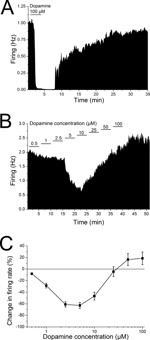
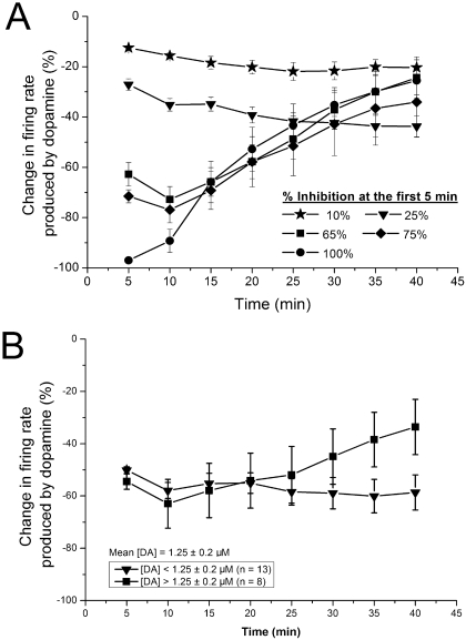
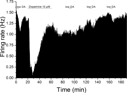
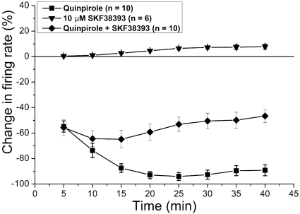
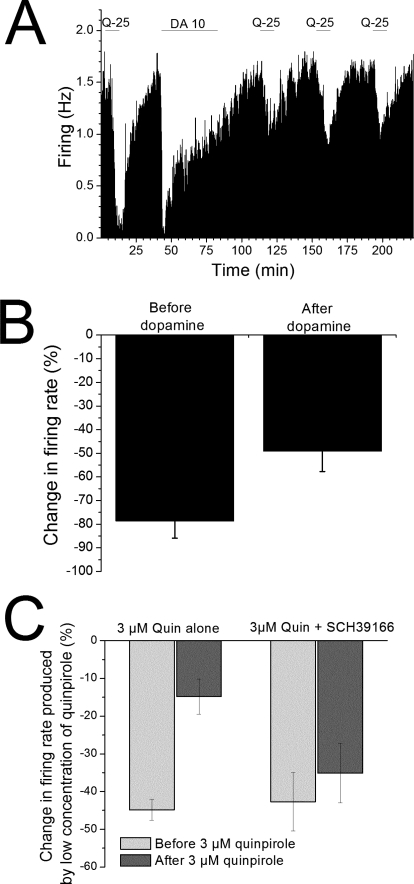
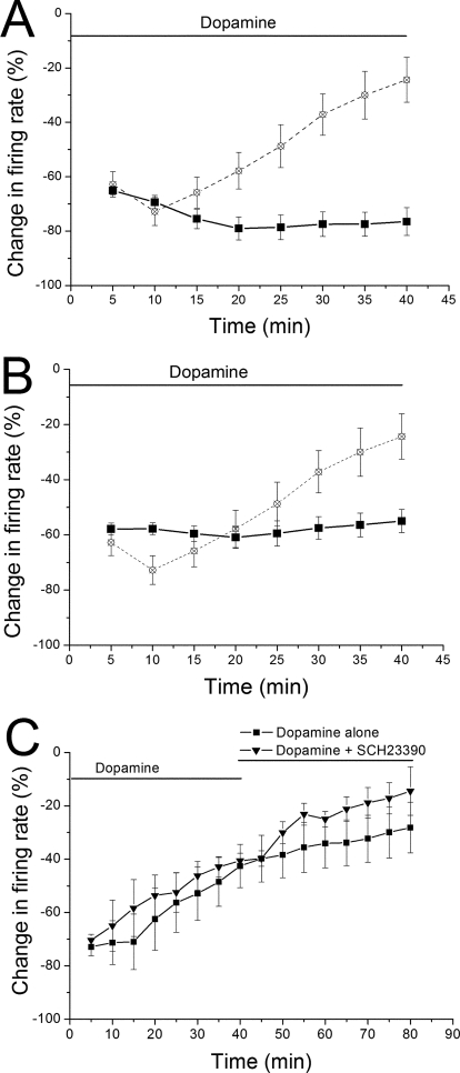
Drug abuse-induced plasticity of putative dopaminergic (pDAergic) ventral tegmental area (VTA) neurons may play an important role in changes in the mesocorticolimbic system that lead to the development of addiction. In the present study, extracellular recordings were used to examine time-dependent effects of dopamine (DA) on pDAergic VTA neurons in rat brain slices. Administration of DA (2.5-10 microM) for 40 min resulted in inhibition followed by partial or full reversal of that inhibition. The reduced sensitivity to DA inhibition lasted 30 to 90 min after washout of the long-term dopamine administration. The inhibition reversal was not observed with 40-min administration of the D2 agonist quinpirole (25-200 nM), so this phenomenon was not the result of desensitization induced solely by stimulation of D2 DA receptors. Inhibition reversal could be observed with the coapplication of quinpirole and the D1/D5 agonist SKF38393 [(+/-)-1-phenyl-2,3,4,5-tetrahydro-(1H)-3-benzazepine-7,8-diol hydrobromide], suggesting a D1/D5 mechanism for the reversal. Furthermore, D1/D5 antagonists, given in the presence of prolonged DA exposure, prevented the inhibition reversal. Application of 3 microM quinpirole caused desensitization to low quinpirole concentrations that was blocked by a D1/D5 antagonist. These data suggest that coactivation of D1/D5 receptors and D2 receptors in the VTA results in desensitization of autoinhibitory D2 receptors. Prolonged increases in pDAergic tone in the VTA that may occur in vivo with drugs of abuse could reduce the regulation of firing by D2 dopamine receptor activation, producing long-term alteration in information processing related to reward and reinforcement.


Figures

Fig. 1.
Fig. 1.
Dopamine concentration-response curves using stepwise application of a range of dopamine concentrations. A, rate-meter graph of the effect of acute application of a high DA concentration. Vertical bars indicate the firing rate over 5-s intervals. Horizontal bars indicate the duration of application of DA (100 μM). Administration of DA produced complete cessation of firing, and the firing resumed after washout of the dopamine from the superfusate. B, rate-meter graph of the effect of stepwise application of a series of DA concentrations (0.5–100 μM). Vertical bars indicate the firing rate over 5-s intervals. Horizontal bars indicate the duration of application of DA (concentration in micromolar indicated above bar). Note that even though the concentration of dopamine was increased every 5 min, inhibition only increased up to 10 μM dopamine, and as concentrations were increased above 10 μM, the firing rate was less inhibited, and eventually firing increased above the predopamine baseline rate. C, change in spontaneous firing rate (mean ± S.E.M.) in the presence of dopamine is plotted as a function of dopamine concentration (log scale). Dopamine was applied in 5-min steps over a range of concentrations (0.5–100 μM, n = 63). There was a significant effect of concentration on inhibition with concentrations of 2.5 to 10 and 100 μM significantly different from control firing rate.
Fig. 2.
Fig. 2.
Dopamine concentration-response curves using long-duration application of single dopamine concentrations. Change in firing rate (mean ± S.E.M.) produced by dopamine is plotted as a function of time. A, effect of dopamine on spontaneous firing rate is plotted as a function of time. Concentrations of dopamine that produced 10, 25, 65, 75, or 100% inhibition in the first 5 min were applied for 40 min. Choice of the concentration based on initial effect of dopamine controlled for sensitivity of the neurons to the inhibitory effect of dopamine. For concentrations that initially produced 10 (n = 7) or 25% (n = 7) inhibition in the firing rate, there was no significant change in the effect of dopamine over time. For concentrations that initially produced 65 (n = 12), 75 (n = 7), or 100% (n = 11) inhibition in firing rate, there was a significant reduction in the inhibitory effect of dopamine over time, with the last three time points significantly different from the 5-min time point. B, effect of dopamine on spontaneous firing rate is plotted as a function of time for two groups of neurons in which dopamine produced 50% inhibition of firing. In more sensitive cells that were inhibited by 50% by low concentrations of dopamine ([DA] <1.25 μM; n = 13), there was no significant change in the effect of dopamine over time. In less sensitive cells that were inhibited by 50% by higher concentrations of dopamine ([DA] >1.25 μM; n = 8), there was a significant reversal of dopamine inhibition with time, with the last three time points significantly different from the 5-min time point.
Fig. 3.
Fig. 3.
Mean rate-meter graphs of the effect of long-duration dopamine exposure on response to shorter dopamine administration. Mean firing rate over 5-s intervals is plotted as a function of time for cells exposed to dopamine for 5-min intervals before and after a 40-min exposure to 10 μM dopamine. Administration of low concentrations of dopamine (0.42 ± 0.12 μM) for 5 min produced inhibition of firing (28.1 ± 3.8%; n = 6). After these test applications, 10 μM dopamine was applied for 40 min. Inhibition reversal was observed for this 40-min exposure to 10 μM dopamine. After washout of 10 μM dopamine, the effect of brief 5-min exposures to lower concentrations of dopamine (same concentrations for each cell as tested before long-term dopamine exposure) were significantly reduced at 30 min (10.0 ± 4.5%) but was not significantly different at 60 (17.0 ± 1.7%) or 90 min (18.2 ± 1.6%) after the long-term exposure (one-way ANOVA, F = 5.53, p < 0.01; Tukey post-hoc test, P < 0.05 for significance).
Fig. 4.
Fig. 4.
Inhibitory effects of long-duration treatment with quinpirole and SKF38393. Long-duration applications to VTA dopamine neurons of D2 agonist quinpirole, D1/D5 agonist SKF38393, and the combination were performed. Quinpirole (43.5 ± 3.4 nM; n = 10) alone produced significant inhibition (one-way repeated measures ANOVA, P < 0.05; n = 10) that reached a peak at approximately 15 min, and the firing rate remained inhibited for the duration of quinpirole application. SKF38393 (10 μM) alone (n = 6) had a small but significant excitatory effect on the firing rate. In the presence of 10 μM SKF38393, quinpirole (102 ± 20 nM) inhibition partially reversed so that the inhibition at 40 min was significantly less than that at 5 or 10 min (n = 10).
Fig. 5.
Fig. 5.
Effect of quinpirole after induction of dopamine-inhibition reversal. A, rate-meter graph from a single putative dopaminergic neuron. Vertical bars indicate the firing rate averaged over a 5-s interval; horizontal bars indicate the duration of application of either 25 nM quinpirole (Q-25) or 10 μM dopamine (DA-10). Note that the inhibitory effect of quinpirole after the 40-min dopamine treatment is much less than that observed before the dopamine treatment. Initially, quinpirole produced inhibition of approximately 90%; after dopamine treatment, the quinpirole-induced inhibition (tested at 30-min intervals) was 25, 45, and 44%, respectively. B, effect of quinpirole before and after dopamine in the population of cells tested. Nine cells were tested in a protocol similar to that illustrated in A, and comparison was made between responses to quinpirole before and after dopamine treatment. The mean inhibition produced by quinpirole before dopamine was 78.6 ± 7.3%; 30 min after washout of dopamine, the quinpirole-induced inhibition was 49.1 ± 8.6% (t test, P < 0.02; n = 9). C, effect of quinpirole before and after 3 μM quinpirole. Thirteen cells were tested in a protocol similar to that illustrated in A, with the exception that 3 μM quinpirole was applied for 25 min instead of the 40-min dopamine application. Comparison was made between responses to low concentrations of quinpirole before (light bar) and after (dark bar) 3 μM quinpirole treatment. The mean inhibition produced by low quinpirole (mean concentration = 36 ± 13 nM) before the 3 μM application was 44.8 ± 2.8%; within 1 h of the washout of 3 μM quinpirole, the same concentration of quinpirole induced an inhibition of only 14.8 ± 4.7% (t test, P < 0.005; n = 6). In the presence of 10 μM SCH39166, the mean inhibition produced by low quinpirole (mean concentration = 285 ± 77 nM) before the 3 μM application was 42.7 ± 7.8%; within 1 h of the washout of 3 μM quinpirole, the same concentration of quinpirole induced an inhibition of 35.1 ± 7.9% that was not significantly different from the value before 3 μM quinpirole treatment (t test, P > 0.05; n = 7).
Fig. 6.
Fig. 6.
Effects of D1/D5 antagonists on dopamine-inhibition reversal. A and B, antagonists applied 10 min before dopamine: a concentration of dopamine sufficient to cause inhibition of 60% or greater was applied for 40 min in the presence of either 10 μM SCH39166 (dopamine concentration = 2.4 ± 0.4 μM; n = 10) (A) or 10 μM SCH23390 (dopamine concentration = 3.9 ± 1.4 μM; n = 8) (B). No significant reduction in dopamine-induced inhibition was observed in the presence of either antagonist. On both A and B, the response to a concentration of dopamine that produced 65% inhibition (from Fig. 2) is shown for reference (open symbols and dashed line). C, antagonists applied after dopamine: a concentration of dopamine sufficient to cause inhibition of 60% or greater was applied for 40 min, and then dopamine was continued for an additional 40 min, either alone (n = 6, mean dopamine concentration = 5.1 ± 0.9 μM) or in the presence of 10 μM SCH23390 (n = 4, dopamine concentration = 4.0 ± 1.4 μM). There was a significant reduction in the effect of dopamine over the 80-min period in both groups (two-way ANOVA, F = 7.37, p < 0.001) and a statistically significant difference in the effect of dopamine overall between the cells treated with dopamine alone (n = 6) or with dopamine plus SCH23390 (n = 4) (two-way ANOVA, p < 0.02). Note that there was no antagonism of the dopamine-inhibition reversal with the addition of SCH23390.

Similar articles

Cited by

References

    1. Argilli E, Sibley DR, Malenka RC, England PM, Bonci A. (2008) Mechanism and time course of cocaine-induced long-term potentiation in the ventral tegmental area. J Neurosci 28:9092–9100 - PMC - PubMed
    1. Bartlett SE, Enquist J, Hopf FW, Lee JH, Gladher F, Kharazia V, Waldhoer M, Mailliard WS, Armstrong R, Bonci A, et al. (2005) Dopamine responsiveness is regulated by targeted sorting of D2 receptors. Proc Natl Acad Sci U S A 102:11521–11526 - PMC - PubMed
    1. Barton AC, Black LE, Sibley DR. (1991) Agonist-induced desensitization of D2 dopamine receptors in human Y-79 retinoblastoma cells. Mol Pharmacol 39:650–658 - PubMed
    1. Brodie MS, Dunwiddie TV. (1987) Cholecystokinin potentiates dopamine inhibition of mesencephalic dopamine neurons in vitro. Brain Res 425:106–113 - PubMed
    1. Brodie MS, Dunwiddie TV. (1990) Cocaine effects in the ventral tegmental area: Evidence for an indirect dopaminergic mechanism of action. Naunyn Schmiedebergs Arch Pharmacol 342:660–665 - PubMed

Publication types

MeSH terms

LinkOut - more resources